Are you sure you want to perform this action?
Appendectomy, laparoscopic
You have not purchased a license - paywall is active: to the product selection
Single Access
Access to this lecture
for 3 days
€4.99 inclusive VAT

webop-Account Single
full access to all lectures
price per month
for the modul: vascular surgery
from 8,17 €
-
Relevant surgical anatomy
Surgical anatomy of the right lower quadrant
The colon is about 1.5m long and originates at the confluence of the small intestine into the cecum. The cecum is located inferior to the ileocecal valve, has its own mesentery (→ mobility) with the appendicular artery and vein (← ileocolic artery← superior mesenteric artery) and is approx. 7cm long. The cul-de-sac tubular vermiform appendix arises from the medial wall of the cecum directly caudad of the ileocecal valve in the taenia libera. It is intraperitoneal, with a length of 2–20cm and a diameter between 0.5cm and 1cm. Usually, the appendix extends from the posterior middle of the cecum to the center of the body, but its position can be quite variable, and thus also the location and severity of tenderness. This surgical condition is incorrectly referred to as "appendicitis", although from an anatomical point of view the affected organ is merely the vermiform appendix of the cecum.
Anatomical variants of the appendix:
- Descending type: Appendix extending into the lesser pelvis. In women it may be closely adjacent to the ovary.
- Medial position: Appendix between loops of the small intestine.
- Lateral position: Appendix between the lateral abdominal wall and cecum.
- Retrocecal position: Appendix turned craniad posterior to the cecum (65%)
- Anterocecal position: Appendix turned craniad anterior to the cecum.
- Subhepatic position: Appendix turned toward the liver and in contact with it.
Histologically, the mucous membrane of the appendix demonstrates the same makeup as in the large bowel. However, it has a large number of lymphatic cells and thus becomes part of the human immune system. In addition, in the wall of the appendix the 3 individual strips of longitudinal muscles (taenia) characteristic of the colon conjoin to a complete layer once again.
Follow this link for further information on the anterior abdominal wall.
Single Access
Access to this lecture
for 3 days
€4.99 inclusive VAT

webop-Account Single
full access to all lectures
price per month
for the modul: vascular surgery
from 8,17 €
Single Access
Access to this lecture
for 3 days
€4.99 inclusive VAT

webop-Account Single
full access to all lectures
price per month
for the modul: vascular surgery
from 8,17 €
-
Instruments, draping, minilaparotomy
-
Pneumoperitoneum, exploration
Establish the pneumoperitoneum.
The pressure level should reflect size, age and sex of patient (6–8mmHg in children, 10–14mmHg in adults). Limit the CO2flow rate to 1mL/min.Abdominal exploration.
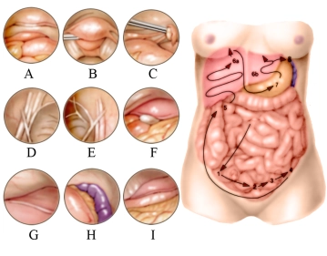
The video demonstrates acute appendicitis. Laparoscopy always starts with the abominal exploration! In the video, first the right lower quadrant is explored, then the right upper quadrant, left upper quadrant, again the area of the appendix, and finally the lesser pelvis. The lower abdominal organs are inspected in the numerical order of the sketch: Inspection around the appendix is followed by uterus and bladder (A), Douglas pouch (B), left ovary (C), and deep inguinal orifice (D). This is continued to the right groin area (E). Then the laparoscope is directed into the right upper quadrant (5) where the gall bladder (F) and right hepatic lobe (G) are inspected. This is followed by inspection of the left upper quadrant: Spleen and gastric corpus (H) as well as left hepatic lobe with falciform ligament, stomach and greater omentum (I).
-
Working trocars, freeing the cecum
-
Dissecting and resecting the appendix
Bluntly dissect the mesoappendix close to the base of the appendix with Overholt forceps (in video with scissors).
Insert the Endo GIA™ (linear stapler, 30 mm, white cartridge) via the 13.5mm trocar and resect the appendix.
Note: In the video, the"safe” way of resection is taken. Of course, the appendix may also be resected without a linear cutter, e.g., with a Roeder loop. -
Transecting the mesoappendix, irrigation, suction
Transecting the mesoappendix (2ndcartridge).
The mesoappendix may also be transected with diathermy (caution: risk of secondary bleeding).
Only explore for Meckel diverticulum if the appendix appears rather unremarkable.
In Meckel diverticulum:
Resect the diverticulum sufficiently far from its base with a linear cutter (e.g., Endo GIA™ blue 30mm or 45mm cartridge) (not shown in video).
Check for any bleeding. Irrigate and suction the surgical field, particularly the Douglas pouch (evacuate it!). -
Retrieving the appendix
-
Removing the specimen retrieval bag and trocars
-
Fascial suture, skin suture, dressing
Single Access
Access to this lecture
for 3 days
€4.99 inclusive VAT

webop-Account Single
full access to all lectures
price per month
for the modul: vascular surgery
from 8,17 €
Single Access
Access to this lecture
for 3 days
€4.99 inclusive VAT

webop-Account Single
full access to all lectures
price per month
for the modul: vascular surgery
from 8,17 €
-
Literature summary
-
Ongoing trials on this topic
-
References on this topic
-
literature search
Literature search under: http://www.pubmed.com