Start your free 3-day trial — no credit card required, full access included

Anatomy - Left hemicolectomy, open, curative

  1. Overview

    Overview
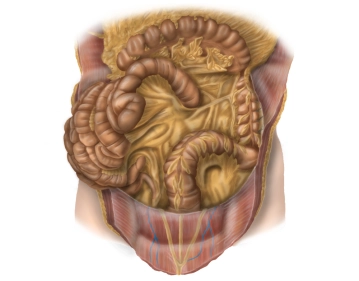
    • The colon surrounds the loops of the small intestine along the inner abdominal wall and below the liver and stomach. The position of the colon is intra- or secondarily retroperitoneal. Its primary function is the thickening of the chyme through the absorption of water. The total length of the colon is on average 120-150 cm. The colon begins at the ileocecal valve and ends at the rectosigmoid junction, where it transitions into the rectum.
    • The colon is divided into the following sections:
      • Cecum with the appendix
      • Ascending colon
      • Transverse colon
      • Descending colon
      • Sigmoid colon
  2. Macroscopy

    Macroscopy
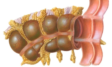
    • The longitudinal muscle layers of the colon form three band-like muscle strips that are visible from the outside and are referred to as taeniae. They are distinguished as follows:
      • Taenia mesocolica: located towards the mesentery
      • Taenia libera: free on the surface, facing the abdominal wall
      • Taenia omentalis: connected with the greater omentum
    • Appendices epiploicae are fat appendages from the tela subserosa in the area of the free taeniae.
    • Plicae semilunares are indentations of all wall layers intraluminally, whereas haustra are the outward bulges in between.
    • For left hemicolectomy, the following intestinal segments are relevant from oral to aboral:
      • distal transverse colon
      • left flexure
      • descending colon
      • sigmoid colon
      • upper rectum
  3. Transverse colon, descending colon, and sigmoid colon

    Transverse colon, descending colon, and sigmoid colon
    • Transverse colon
      • Synonyms: transverse colon or simply Transversum.
      • Location: intraperitoneal; mobile fixation through its own mesentery – transverse mesocolon, runs transversely ascending through the abdominal cavity from the right to the left flexure
      • Length: 30-45 cm
      • Identification: by the three taeniae and the greater omentum attached to the taenia omentalis
      • The left flexure is always higher than the right flexure due to its fixation by the phrenocolic ligament
    • Descending colon:
      • Location: secondarily retroperitoneal
      • Peritoneal suspension: fused with the posterior wall of the abdominal cavity
      • Course: from the left flexure (here: phrenocolic ligament with fixation to the spleen) to the iliac fossa, connects to the transverse colon and transitions into the sigmoid colon
      • Length: 20-30 cm
    • Sigmoid colon
      • Location: intraperitoneal
      • Peritoneal suspension: sigmoid mesocolon
      • Course: from the iliac fossa as a loop (S-shaped) to the level of the 2nd-3rd sacral vertebrae, connects to the descending colon and transitions into the rectum
      • Length: variable (elongated sigmoid), generally: about 35 cm
  4. Vascular supply, lymphatic drainage, and nerves

    Vascular supply, lymphatic drainage, and nerves
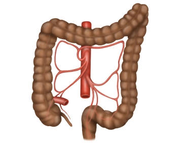
    • Arterial supply of the left hemicolon, sigmoid colon up to the upper rectum by the inferior mesenteric artery.
      • Origin of the inferior mesenteric artery from the abdominal aorta at the level of L3 vertebra.

    Note: Inferior mesenteric plexus (inferior mesenteric ganglion) surrounds the origin of the inferior mesenteric artery.

    • Left colic artery: supplies the descending colon, an ascending branch anastomoses with the middle colic artery, a descending branch anastomoses with a sigmoid artery.
    • Sigmoid arteries: 2-4 arteries, several small branches to the sigmoid colon, anastomoses with the left colic artery and superior rectal artery.
    • Superior rectal artery: runs dorsally to the upper rectum, anastomoses with the sigmoid artery and middle rectal artery from the internal iliac artery.
    • Left colonic flexure: watershed area between the supply territories of the superior mesenteric artery and the inferior mesenteric artery.
      • Anastomosis between the superior mesenteric artery (middle colic artery) and the inferior mesenteric artery (left colic artery) near the left colonic flexure distally (Riolan's anastomosis).

    Note: Inconstant: Riolan's anastomosis is not or not sufficiently developed in 20% of cases.

    • Drummond's arcade: vascular arcade that connects the branches of the colic branches of the superior mesenteric artery and the inferior mesenteric artery near the intestine and runs caudally close to the colon.
    • Venous drainage via the left colic vein and sigmoid veins into the inferior mesenteric vein, which drains into the splenic vein behind the tail of the pancreas. This forms the portal vein confluence with the superior mesenteric vein and other visceral veins behind the head of the pancreas.
    • Lymphatic drainage along the course of the inferior mesenteric artery (left colic lymph nodes, sigmoid lymph nodes).
    • Important neural structures:
      • Inferior mesenteric plexus (inferior mesenteric ganglion) (autonomic nervous system) at the origin of the inferior mesenteric artery.
      • Involved sympathetic nerves: lumbar splanchnic nerves, involved parasympathetic nerves: pelvic splanchnic nerves via the inferior hypogastric plexus.
  5. Topographical relationships

    Topographical relationships
    • The transverse colon runs across the abdomen from right to left, slightly ascending, and has close anatomical relationships with:
      • Stomach (connected by the gastrocolic ligament)
      • Greater omentum
      • Anterior surface of the pancreas
      • Lesser sac
    • The descending colon runs from cranial to caudal adjacent to the left lateral abdominal wall. It has a close anatomical relationship with:
      • Spleen (left colic flexure), 
      • Lesser sac (dorso-medial to the left colic flexure), 
      • Left kidney
      • Tail of the pancreas (cranial section of the descending colon).

    Note: The left colic flexure is fixed to the diaphragm by the phrenicocolic ligament and to the spleen by the splenocolic ligament

    • The sigmoid colon runs in an S-shape from the left iliac fossa into the pelvis. The root of the sigmoid mesocolon crosses from the lateral iliac fossa medially:
      • the common iliac vessels, 
      • the ureter
      • the ovarian or testicular vessels.

    Note: Course of the ureter:

    • dorsal to the descending colon and sigmoid colon under Gerota's fascia
    • on the psoas muscle, which it crosses from latero-cranial to caudo-medial