Are you sure you want to perform this action?
Intersphincteric resection, open, with transanal colon pull-through and transverse coloplasty pouch
You have not purchased a license - paywall is active: to the product selection
Single Access
Access to this lecture
for 3 days
€4.99 inclusive VAT

webop-Account Single
full access to all lectures
price per month
for the modul: vascular surgery
from 8,17 €
-
Descending and sigmoid colon
- Splenic flexure attached to the diaphragm by the phrenicocolic ligament
- Descending colon situated in secondary retroperitoneal position
- Transition between descending and sigmoid colon in the left iliac gutter
- Sigmoid colon situated intraperitoneally (→ sigmoid mesocolon)
- Transition between sigmoid colon and rectum anterior to the 2nd/3rdsacral vertebra
-
Rectum
The rectum is divided into thirds. The level of their borders is measured with the rigid endoscope and referenced to the anocutaneous line. Distal third at 0-6cm, middle third at 6-12cm, proximal third at 12-16cm
-
Fascia systems
- The pelvic parietal fascia covers the pelvic wall with its vessels, autonomic nerves and plexus of the presacral veins/nerves
- The rectosacral fascia and visceral pelvic fascia meet proximal to the anorectal transition
- The visceral fasciae comprise the proper pelvic fascia (encasing the posterior and lateral mesorectum) and the anterior Denonvilliers fascia
-
Vessels
- Anastomoses between branches of the superior and inferior mesenteric artery (middle and left colic artery respectively) near the splenic flexure (anastomosis of Riolan).
- Left hemicolon, sigmoid colon and upper rectum supplied by the inferior mesenteric artery and its branches: left colic artery, sigmoid arteries with Drummond marginal artery, superior rectal artery.
- Proximal third of the rectum supplied by the superior rectal artery dividing posteriad into two terminal branches; middle third by the middle rectal artery (paired, each originating from the internal iliac artery); and the distal third by the inferior rectal artery (paired, each originating from the internal iliac artery). The middle rectal arteries course in the so-called lateral ligaments of the rectum and are divided in total mesorectal excision.
- Venous blood from the left hemicolon flows via veins of the same name into the area drained by the portal vein.
- Venous drainage of the proximal and middle third via the inferior mesenteric vein (draining into the portal vein), while venous blood from the distal third returns via the area drained by the inferior vena cava.
-
Lymphatic drainage
- For all rectal segments along the course of the superior rectal artery and inferior mesenteric artery.
- In the distal third also via lymphatics paralleling the middle rectal and internal iliac arteries to lymph nodes around the levator ani muscles and ischioanal fossa.
-
Topographical relations
Descending colon, retroperitoneum and rectum
- Course of the descending colon from proximal to distal closely related to the spleen (splenic flexure), omental bursa (posteromedial to the splenic flexure), left kidney and pancreatic tail (proximal segment of descending colon).
- Root of the sigmoid mesocolon from the left iliac gutter coursing mediad, crossing the common left iliac vessels, left ureter and left ovarian/testicular vessels.
- Left ureter coursing on the psoas muscle, which it crosses from superolaterad to inferomediad.
-
Lateral rectal topography
Single Access
Access to this lecture
for 3 days
€4.99 inclusive VAT

webop-Account Single
full access to all lectures
price per month
for the modul: vascular surgery
from 8,17 €
Single Access
Access to this lecture
for 3 days
€4.99 inclusive VAT

webop-Account Single
full access to all lectures
price per month
for the modul: vascular surgery
from 8,17 €
-
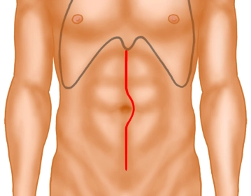
Skin incision and exposure
-
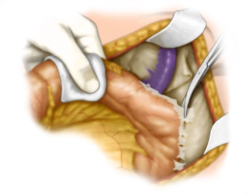
Freeing the left colon
-
Managing the inferior mesenteric vein
-
Managing the inferior mesenteric artery
-
Entering the lesser pelvis
After instituting Trendelenburg position, open the peritoneum at the base of the mesosigmoid and mesorectum on both sides from anteriorly to the promontory down to the pouch of Douglas, and then open the "holy plain" posterior to the mesorectum, respecting the layers and the hypogastric nerves.
Perform this dissection akin to total mesenteric excision. -
Mesorectal dissection
-
Anterior rectal dissection
Dissect both lateral ligaments and divide the ligament structures between the coccyx and rectum. Now incise the anterior peritoneal reflection between the bladder /uterus and rectum and dissect down to the Denonvilliers fascia. While staying within the correct layer carry the dissection down to the suprasphincteric/intersphincteric level. In men take particular care to respect the plexus of nerves and veins encasing the seminal vesicles. The inferior hypogastric plexus on the anterolateral pelvic floor must be spared.
Tip: In women, dissection takes place in the retrovaginal space. In anterior cancer, partial resection of the posterior vaginal wall may be required.
-
Rectal palpation
-
Exposing the puborectalis muscle
-
Mounting the Lone Star Retractor System™
-
Submucosal injection for hemostasis
-
Intersphincteric excision
-
Delivering the resectate
-
Pouch construction
-
Preplacing the interrupted sutures for the anastomosis
-
Pulling the oral stump through the lesser pelvis
-
Completing and tying the interrupted sutures
-
Removing the anal retractor
Single Access
Access to this lecture
for 3 days
€4.99 inclusive VAT

webop-Account Single
full access to all lectures
price per month
for the modul: vascular surgery
from 8,17 €
Single Access
Access to this lecture
for 3 days
€4.99 inclusive VAT

webop-Account Single
full access to all lectures
price per month
for the modul: vascular surgery
from 8,17 €
-
Zusammenfassung der Literatur
-
Aktuell laufende Studien zu diesem Thema
-
Literatur zu diesem Thema
-
Reviews
-
Guidelines
-
literature search
Literature search under: http://www.pubmed.com