Heterotopic parathyroid autotransplantation in total parathyroidectomy - general and visceral surgery

You have not purchased a license - paywall is active: to the product selection
  • Kocher or collar incision

    Video
     
    Kocher or collar incision
     

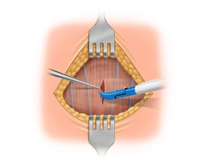
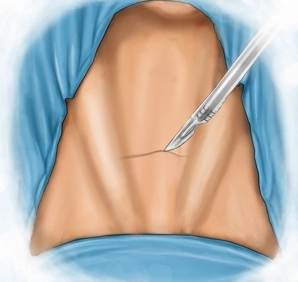
    Standard skin incision 2 finger widths (2-3 cm) superior to the suprasternal notch between the bellies of the left and right sternocleidomastoid muscles.

    With the Ligasure® divide subcutaneous tissue and platysma down to anterior fascia of the neck.

    Expose the avascular plane between platysma and anterior cervical fascia of and free the skin-platysma flap cephalad and caudad.

  • Midline division of the strap muscles

    Video
     
    Midline division of the strap muscles
     

    Divide the fascia and strap muscles in the midline while sparing the superficial veins of the neck. Then retract the strap muscles of the neck from the anterior aspect of the thyroid.

  • Freeing the right superior pole of the thyroid

    Video
     
    Freeing the right superior pole of the thyroid
     

    Turn to the right thyroid lobe. Dissect the right side and successively free the superior pole. Expose the vessels of the superior pole and transect them close to the lobe between clips and Ligasure® seals.

  • Exposing the vagus nerve

    Video
     
    Exposing the vagus nerve
     

    Carefully free the right thyroid lobe while transecting the medial thyroid vein (Kocher vein).

    Locate and underrun the vagus nerve with a vessel loop. Neuromonitoring with the Medtronic system demonstrates normal signals.

  • Resection of the superior right parathyroid

    Video
     
    Resection of the superior right parathyroid
     

    Locate the recurrent laryngeal nerve and verify it by neuromonitoring. Then expose the superior right parathyroid at its typical location posterior to the recurrent laryngeal nerve.

    After sealing with the Ligasure® resect the parathyroid between clips.

  • Exploring the inferior parathyroid at its usual location

    Paid content (video)
    Paid content (image)

    After resection of the enlarged nodular right thyroid lobe (not illustrated), once more locate and neuromonitor the recurrent laryngeal nerve, visible in its entire course,

    Then dissect at the usual location of the inferior parathyroid anterior to the nerve and inferior the inferior thyroid artery.

    Since the parathyroid cannot be located here, continue the dissection along the thyrothymic ligament to the right lobe of the thymus.

  • Starting the right transcervical thymectomy

    Paid content (video)
    Paid content (image)

    Step by step and under gentle traction free the thymus cephalad outside its delicate capsule. Finally, within the right thymic lobe a markedly enlarged parathyroid is seen which can be resected after placing an inferior clip.

  • Thymectomy

    Paid content (video)
    Paid content (image)

    Precise anterior dissection between clips and Ligasure® seals avoids bleeding and spares the recurrent laryngeal nerve.

    And finally, the left lobe of the thymic is also resected and extracted cephalad after deep ligature directly adjacent to the brachiocephalic trunk. The third parathyroid gland is now seen within the left lobe of the thymus.

  • Freeing the left thyroid lobe

    Paid content (video)
    Paid content (image)

    Transect the right Kocher vein and free the left thyroid lobe laterally. Pull the lobe in an anteromedial direction with atraumatic Babcock forceps. Locate the vagus nerve within the carotid sheath and underrun it with a vessel loop. Verify correct functioning of the recurrent laryngeal nerve by neuromonitoring. After opening the space between the posterolateral aspect of the thyroid and the carotid sheath, filled with delicate alveolate connective tissue, expose the inferior thyroid artery. The recurrent laryngeal nerve crosses here and can be verified by neuromonitoring. After clip placement transect the left thyrothymic ligament and remove the entire thymus.

  • Resection of the superior left parathyroid

    Paid content (video)
    Paid content (image)

    The left superior parathyroid gland manifests in its characteristic location superior to the inferior thyroid artery and is resected after sealing the delicate vessels with the Ligasure® forceps.

  • Final neuromonitoring

    Paid content (video)
    Paid content (image)
    Paid content (text)
  • Drain insertion

    Paid content (video)
    Paid content (image)
    Paid content (text)
  • Layered wound closure

    Paid content (video)
    Paid content (image)
    Paid content (text)
  • Preparing the graft

    Paid content (video)
    Paid content (image)
    Paid content (text)
  • Autotransplantation

    Paid content (video)
    Paid content (image)
    Paid content (text)