Start your free 3-day trial β€” no credit card required, full access included

Right adrenalectomy, open

Reading time readingtime 28:02 min.
  1. Laparotomy; Exploration

    Video
    Laparotomy; Exploration
    Soundsettings

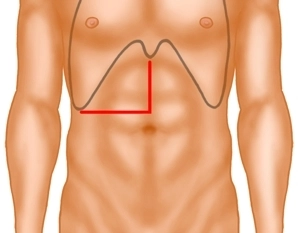
    The laparotomy begins with a right-sided transverse incision at the level of the navel and is then extended in the midline supraumbilically to the xiphoid. Examination of the abdominal organs to exclude further metastases, confirmation of resectability.

  2. Mobilization of the Duodenum according to Kocher

    Video
    Mobilization of the Duodenum according to Kocher
    Soundsettings

    The liver is elevated and the duodenum is mobilized and retracted medially to such an extent that the inferior vena cava is accessible in its full length.

    Note: Detachment of the right colonic flexure may additionally be necessary depending on the operating field (not required in this example).

  3. Depiction of the Right Retroperitoneum with Cranial and Medial Displacement of the Liver

    Video
    Depiction of the Right Retroperitoneum with Cranial and Medial Displacement of the Liver
    Soundsettings


    To achieve a sufficiently good depiction of the right adrenal gland, which is retroperitoneal and located directly under the liver next to the inferior vena cava, it is necessary to displace the liver cranially and medially. First, the parietal peritoneum covering the front of the lower part of the adrenal gland is opened parallel to the inferior vena cava. 

    Note: Surgical procedures on the right retroperitoneum are more challenging than on the left side, as liver enlargement, obesity, or a barrel chest can significantly complicate the positioning of the surgical site.

  4. Mobilization of the liver from caudal; preparation at the inferior vena cava

    Video
    Soundsettings

    Next, the adhesions between the liver surface and the adrenal gland are severed. Due to the peritumoral inflammatory reaction, there is close contact here.

    Then preparation of the retrohepatic inferior vena cava, with individual veins draining into segment 1 being severed to allow the right liver lobe to be displaced far enough to the left.


  5. Detachment of the Adrenal Gland from the Kidney

    Video
    Detachment of the Adrenal Gland from the Kidney
    Soundsettings

    Now the adrenal tumor is detached from the upper pole of the kidney and from the surrounding perirenal fat tissue.

  6. Dissection of the Adrenal Vein

    Video
    Dissection of the Adrenal Vein
    Soundsettings

    The most important landmark is the inferior surface of the liver and the lateral edge of the vena cava. Dissection along the lateral edge of the vena cava leads to the identification of the right adrenal vein. Venous drainage from the adrenal gland typically occurs through a single vein, the suprarenal vein. It exits the glandular tissue at the anterior surface of the adrenal gland. The right adrenal vein drains directly into the inferior vena cava after a short course.

  7. Retro-caval Dissection of the Adrenal Tumor

    Video
    Retro-caval Dissection of the Adrenal Tumor
    Soundsettings

    Then the dissection continues behind the vena cava. Here, the arterial blood supply is encountered, which can be transected after placing clips.

    Note: The numerous smaller arterial branches from the aorta and renal artery are of minor importance.

  8. Removal of the Specimen

    Video
    Removal of the Specimen
    Soundsettings

    For large tumors, looping the renal vein is helpful for orientation. This is followed by the stepwise mobilization of the adrenal tumor in the retroperitoneum dorsal to the vena cava. The specimen is finally excised en bloc.

  9. Diaphragm Suture; Liver Adhesion

    Video
    Diaphragm Suture; Liver Adhesion
    Soundsettings

    After tumor-related necessary inclusion of diaphragm parts, it is necessary to close the diaphragm defect with a continuous suture. The decapsulated liver surface that arises with large adrenal tumors is checked for bleeding and bile leakage, and covering with a hemostatic sealing matrix is recommended. Smaller lesions are coagulated with the argon beam.

  10. Drainage; Abdominal Wall Closure

    Video
    Soundsettings

    After placing a drainage in the right adrenal bed, the abdominal wall closure is performed with a two-layer fascial closure and an intracutaneous suture.

Adverts

Active local hemostasis and sealing

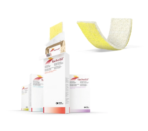
TachoSil® Versiegelungsmatrix

TachoSil® is used in adults and children from 1 month of age as supportive treatment in surgery for improving hemostasis, for supporting tissue sealing, and for suture support in vascular surgery when standard techniques are insufficient. TachoSil® is used in adults for supportive sealing of the dura mater to prevent postoperative cerebrospinal fluid leakage after neurosurgical procedures.

Produktwebsite TachoSil®
TachoSil® Prescribing Information 05-2025 (354.1 kB)

to top