Right adrenalectomy, open

Reading time readingtime 11:49 min.
  1. Laparotomy and exploration

    Video
    Laparotomy and exploration

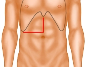
    Transverse right laparotomy at the level of the umbilicus. After transecting the right rectus muscle with bipolar scissors and opening the peritoneum extend the incision in the midline to the xyphoid process. Explore the intestinal organs to rule out other metastases and confirm resectability.

  2. Kocher maneuver mobilizing the duodenum

    Video
    Kocher maneuver mobilizing the duodenum

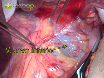
    Depending on the conditions in the field the right colic flexure may have to be freed. Follow this by mobilizing the descending duodenum until the inferior vena cava is exposed along its full length.

  3. Exposing the right retroperitoneum and mobilizing the liver

    Video
    Exposing the right retroperitoneum and mobilizing the liver

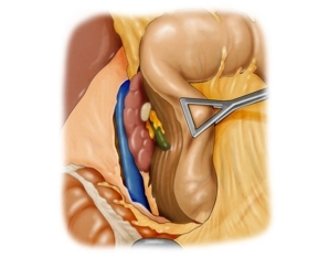
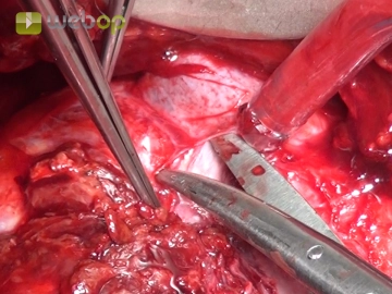
    The posterior aspect the right adrenal abuts the diaphragm. The inferior aspect contacts the right kidney and the anteriosuperior aspect borders on the nude area of the liver. The anteromedial aspect of the right adrenal is posterior to the inferior vena cava. Paralleling the inferior vena cava open up the parietal peritoneum covering the anteroinferior aspect of the right adrenal.

    Transect the right triangular ligament before retracting the liver craniad. Now transect the adhesions between the inferior aspect of the liver and the adrenal. The perineoplastic inflammatory reaction will have resulted in intimate contact.

    Note: Surgical procedures on the right retroperitoneum are more difficult than on the left side because enlargement of the liver, obesity and barrel chest may make visualization and control of the surgical field quite difficult.

  4. Exposing the inferior vena cava

    Video

    Also expose the posterohepatic segment of the inferior vena cava when mobilizing the liver from caudad as well; in doing so, individual veins draining into liver segment 1 must be transected before the right hepatic lobe can be retracted far enough to the left.

  5. Separating right adrenal and kidney

    Video
    Separating right adrenal and kidney

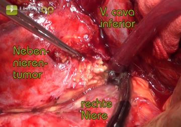
    Now dissect the adrenal tumor off the superior renal pole and its perirenal fatty tissue.

  6. Transecting the adrenal vein

    Video
    Transecting the adrenal vein

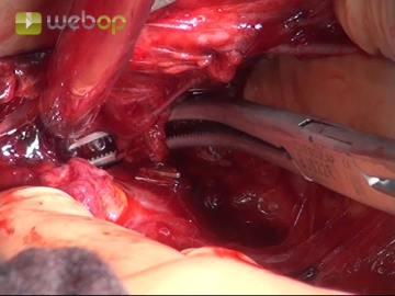
    The most important landmarks are the inferior aspect of the liver and lateral margin of the inferior vena cava. Dissection along the lateral margin of the inferior vena cava will lead to the right adrenal vein.

    Usually, only via a single vein drains the blood from the right adrenal into the suprarenal vein. This vein exits the adrenal at the anterior aspect of the gland. The right adrenal vein courses just a small distance before it drains directly into the inferior vena cava.

  7. Posterocaval dissection of the adrenal tumor

    Video
    Posterocaval dissection of the adrenal tumor

    Now carry the dissection posterior to the inferior vena cava. Here, the arterial blood supply is encountered which is transected between clips.

    Note: The numerous small arterial feeders from the aorta and renal artery are of minor importance.

  8. Specimen removal

    Video
    Specimen removal

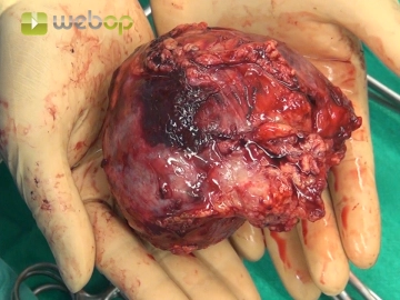
    Orientation in the presence of large tumors is facilitated by encircling the renal vein with a vessel loop. Follow this by gradually dissecting the adrenal tumor off the retroperitoneum posterior to the inferior vena cava. As final step remove the specimen in toto from the surgical field.

  9. Closing the diaphragm and sealing the liver

    Video
    Closing the diaphragm and sealing the liver

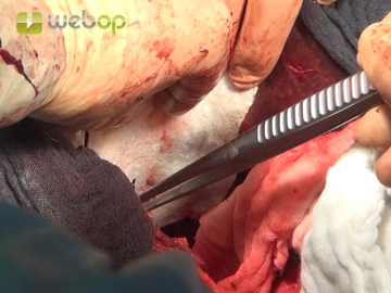
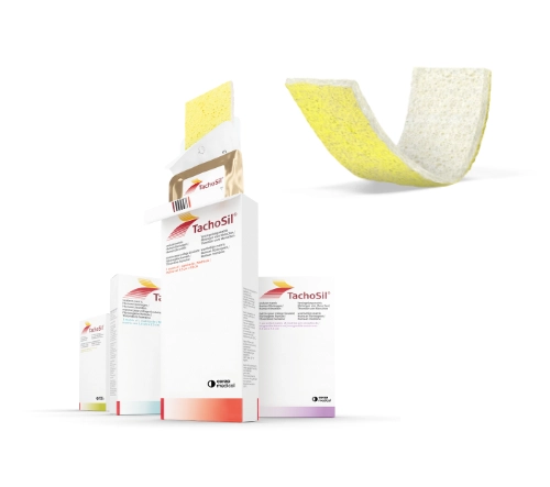
    If tumor resection required partial removal of the diaphragm, close this defect with a running suture. In large adrenal tumors check the decapsulated surface of the liver for leakage of blood and bile; sealing such defects with a TachoSIl® patch is recommended.

  10. Drainage and closing the abdominal wall

    Video

    After draining the right adrenal compartment close the abdominal wall with two-layered suture of the fascia and the skin with a subcuticular suture.

Adverts

Active local hemostasis and sealing

TachoSil® Versiegelungsmatrix

TachoSil® wird bei Erwachsenen und Kindern ab einem Alter von 1 Monat zur unterstützenden Behandlung in der Chirurgie zur Verbesserung der Hämostase, zur Unterstützung der Gewebeversiegelung und zur Nahtsicherung in der Gefäßchirurgie angewendet, wenn Standardtechniken insuffizient sind. TachoSil® wird bei Erwachsenen zur unterstützenden Versiegelung der Dura mater angewendet, um postoperative Liquorleckagen nach neurochirurgischen Eingriffen zu vermeiden.

Produktwebsite TachoSil®
TachoSil® Fachinformation 03-2023 (344.4 kB)