Start your free 3-day trial — no credit card required, full access included

Anatomy - Liver cyst deroofing, laparoscopic

  1. Functional Liver Anatomy

    Functional Liver Anatomy

    Through the falciform ligament and the insertion of the round ligament of the liver on the diaphragmatic as well as the sagittal fissure on the visceral surface, the liver is macroscopically divided into a larger right and a smaller left lobe (volume ratio approx. 80 : 20), although this morphological division does not correspond to the functional structure of the liver. The functional division of the liver is determined by the branching of the portal structures: portal vein, hepatic artery and bile duct. These three anatomical structures branch not only in the porta hepatis, but also within the parenchyma predominantly in the same direction. Each liver segment is completely independent of the other segments in terms of blood supply and bile drainage and can be surgically removed without endangering the function of the remaining liver.

    The term “functional anatomy” thus refers to a substructuring of the liver, which is based on the delimitability of hemodynamically independent parenchyma districts and whose knowledge is essential for the operative strategy in liver resection procedures.

  2. Portal Vein and Hepatic Veins

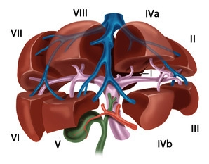
    The functional division of the liver is based on the portal branching into individual, independent subunits, the segments.

    Usually, the portal vein divides in the hepatic hilum into a right and left main trunk. The boundary of these supply areas lies in the cava-gallbladder line (“Cantlie line”). Through renewed bifurcation of the respective portal vein trunk, an anteromedial as well as a posterolateral trunk arises on the right side for the liver segments V/VIII and VI/VII, respectively. The left main trunk extends transversely to the left and then as the pars umbilicaris anteriorly and ends at the insertion site of the ligamentum teres hepatis in the so-called recessus rex. The left portal main trunk gives off branches for the two left-lateral segments II and III as well as for the median segments IVa and IVb. The caudate lobe occupies a special position, as it can receive strong inflows from the left and also from the right portal vein main trunk.

    According to Couinaud, eight portal venous liver segments are distinguished, which, starting with the caudate lobe as segment I, are numbered clockwise:

    Segment I

    Caudate lobe

    Segments I/II/III

    Lateral left hepatic lobe

    Segment IV

    Left paramedian sector (quadrate lobe)

    Segments I/II/III/IV

    Left half of the liver

    Segments V/VIII

    Right paramedian sector

    Segments VI/VII

    Right lateral sector

    Segments V/VI/VII/VIII

    Right half of the liver

    The liver is traversed in a caudocranial direction by three main venous trunks, namely the right, middle, and left hepatic veins, which divide the liver into a total of four hepatic sectors. The left hepatic vein drains almost exclusively the left-lateral hepatic lobe and usually unites shortly before its entry into the vena cava with the middle hepatic vein, which runs along the cava-gallbladder line. The right hepatic vein runs between the posterolateral and anteromedial segments. The caudate lobe has an independent venous drainage, which consists of multiple small veins emptying dorsally directly into the vena cava, the so-called Spieghel veins.

    The portal hila of liver segments II, III, and IV are located extrahepatically and can be relatively easily dissected in the anterior section of the left umbilical fissure. The hila of the right-sided liver segments are located intrahepatically. Exceptions occur occasionally and usually concern segment VI. The anatomy of the hepatic veins is even more variable than that of the portal vein.
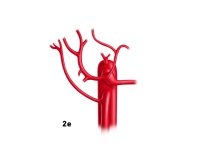
    Variants

    Portal Vein System

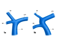
    • Anomalies of the portal vein bifurcation almost always affect the right portal vein main trunk
    • Portal vein trifurcation: right main trunk is absent, instead there are two branches for the right double segments V/VIII and VI/VII (Fig. 1a); occasionally one of the right branches can also originate from the left portal vein main trunk (Fig.1b)
    • Variants of the left portal vein system rarely affect the main trunk, but almost always the division: several small portal vein branches instead of two segmental branches IVa/IVb, occasionally also an additional, intermediate branch between the segmental branches II and III
    44_LAV_01_a_b_Pfortader_Lebervenen

    Fig. 1a and 1b: PV = portal vein, LPV = left portal vein, RA = right anterior portal vein branch, RP = right posterior portal vein branch
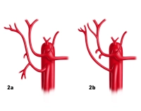
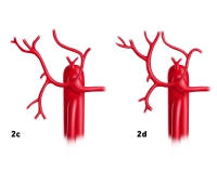
    Hepatic Veins

    • Variants of the hepatic veins are more common than those of the portal vein system
    • Deviations from the hepatic sectors described by Couinaud particularly affect the territories of the right and middle hepatic vein 
  3. Hepatic artery

    The common hepatic artery arises from the celiac trunk; in rare cases, it originates directly from the aorta or the superior mesenteric artery. After giving off the gastroduodenal artery, the proper hepatic artery divides in the hepatic hilum into the right and left hepatic arteries. Not infrequently, additional extrahepatic branches are found, such as the artery for segment IV, which usually arises from the left hepatic artery just before the umbilical fissure.

    Variants
    • Deviations from the normal distribution type are found in 30% of cases
    • the most common anatomical variants are a replaced or accessory hepatic artery from the superior mesenteric artery (Fig. 2a-b) or from the left gastric artery (Fig. 2c-d)
    • occasionally both variants occur together (Fig. 2e)
    44_LAV_02_a_b_Leberarterie
    44_LAV_02_c_d_Leberarterie
    44_LAV_02_e_Leberarterie
Bile Ducts

The extrahepatic portion of the left hepatic duct is approximately 3-5 cm long and arises in the um

Activate now and continue learning straight away.

Single Access

Activation of this course for 3 days.

US$9.50  inclusive VAT

Most popular offer

webop - Savings Flex

Combine our learning modules flexibly and save up to 50%.

from US$7.38 / module

US$88.58/ yearly payment

price overview

general and visceral surgery

Unlock all courses in this module.

US$14.76 / month

US$177.20 / yearly payment

to top