
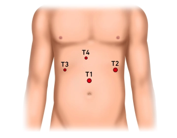
Through a skin incision two to three fingerbreadths above the navel, insertion of the Veress needle and pressure-controlled creation of the pneumoperitoneum up to 13 mm Hg. Insertion of a 10 mm safety trocar. Introduction of the optics and exploration of the upper abdomen: there is an impression of an inflammatory conglomerate tumor; the left liver lobe is adhered cranially to the diaphragm and caudally to the stomach and greater omentum.
-
Creation of the Pneumoperitoneum, Insertion of the Optical Trocar, and Exploration of the Upper Abdomen
![Creation of the Pneumoperitoneum, Insertion of the Optical Trocar, and Exploration of the Upper Abdomen]()
Soundsettings -
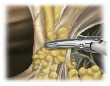
Insertion of the first working trocars and division of the Lig. teres hepatis and the Lig. falciforme
![Insertion of the first working trocars and division of the Lig. teres hepatis and the Lig. falciforme]()
Soundsettings Under endoscopic view insertion of a 12 mm trocar in the left upper abdomen as well as a 5 mm trocar at the same height in the right upper abdomen. The Lig. teres hepatis and the Lig. falciforme hepatis are divided close to the abdominal wall using the UltraCision Harmonic Scalpel®.
-
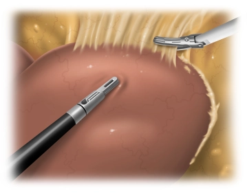
Mobilization of the Liver
![Mobilization of the Liver]()
Soundsettings The mobilization of the left liver lobe is significantly hindered due to inflammatory changes. In this process, adhesions to the diaphragm up to the mobilization of the left hepatic vein must be released step by step. Due to the extensive adhesions, the triangular ligament cannot be identified as such. The stomach and the lesser omentum are also extensively adherent to the visceral surface of the left liver lobe. Detachment of the stomach from the undersurface of the liver with mobilization of the hepatic hilum and opening of the lesser omentum up to the diaphragm.
Local Assessment: Intraoperative Ultrasound (IOUS)
Exchange of the 12 mm upper abdominal trocar on the left for a 15 mm trocar and insertion of the ul
Exchange of the 12 mm upper abdominal trocar on the left for a 15 mm trocar and insertion of the ul
Activate now and continue learning straight away.
Single Access
Activation of this course for 3 days.
US$9.30
inclusive VAT
Most popular offer
webop - Savings Flex
Combine our learning modules flexibly and save up to 50%.
from US$7.23 / module
US$86.85/ yearly payment
general and visceral surgery
Unlock all courses in this module.
US$14.47
/ month
US$173.70 / yearly payment
Webop is committed to education. That's why we offer all our content at a fair student rate.
BiClamp® LAP-Zange
... - Operations in general, visceral and transplant surgery, vascular surgery and thoracic surger
... - Operations in general, visceral and transplant surgery, vascular surgery and thoracic surger
Activate now and continue learning straight away.
Single Access
Activation of this course for 3 days.
US$9.30
inclusive VAT
Most popular offer
webop - Savings Flex
Combine our learning modules flexibly and save up to 50%.
from US$7.23 / module
US$86.85/ yearly payment
general and visceral surgery
Unlock all courses in this module.
US$14.47
/ month
US$173.70 / yearly payment
Webop is committed to education. That's why we offer all our content at a fair student rate.