Start your free 3-day trial — no credit card required, full access included

Inguinal hernia repair using TEP technique

  1. Access to the preperitoneal space

    Video
    Access to the preperitoneal space
    Soundsettings

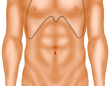
    1 cm long skin incision below the navel. Separate the adipose tissue with Langenbeck hooks and expose the fascia or anterior rectus sheath. This is incised with the scalpel on the side to be treated or, in the case of a bilateral hernia as in the example, on the side of the larger hernia, paramedian over a length of approximately 1 cm. The medial edge of the fascia is grasped with a sharp clamp. Using a Langenbeck hook, the medial rectus muscle is lifted anteriorly and laterally, thus exposing the posterior layer of the rectus sheath.

    Tip: With a precisely median incision of the fascia, one enters directly into the abdomen and not into the rectus sheath. The fascia must then be closed again and incised further laterally!

  2. Insertion of the trocars

    Insertion of the trocars
    Soundsettings

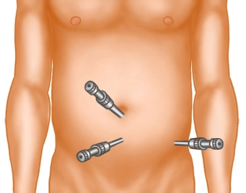
    A long preparation swab is advanced on the posterior leaf towards the symphysis and the preperitoneal space is bluntly dissected up to the retropubic area. Then, a 10mm trocar is inserted and CO2 gas is applied with pressures between 10 – 15 mmHg. The camera optics are introduced. Under visual control, a 5mm trocar is now inserted in the midline exactly between the navel and the symphysis.

    Tip: Before inserting the 5mm trocar, pay attention to the epigastric vessels, which are usually visible. Use trocars with a locking mechanism, as they otherwise slip and significantly complicate the preparation!

  3. Preparation of the preperitoneal space

    Preparation of the preperitoneal space
    Soundsettings

    Through the 5mm trocar, the extraperitoneal space is now mainly bluntly opened with scissors. To do this, begin retrosymphysially and push the tissue from the back of the pubic bone towards the bladder in a ventral to dorsal motion. Small crossing vessels can be coagulated. The retropubic space must be opened below the symphysis.

    Subsequently, dissection is performed laterally in the same manner until the lateral boundary of the rectus sheath appears. This is now incised in tiny steps with scissors until the level of the anterior superior iliac spine is reached. Approximately 3cm above the spine, the second 5mm trocar is inserted under vision.

    Tip: The lateral edge of the rectus sheath should be sharply incised as far ventrally as possible, otherwise, you may enter the abdomen directly!

Preparation of the left side in indirect hernia sac

From now on, use a dissector and fine grasping forceps through the two 5mm trocars. By applying tra

Activate now and continue learning straight away.

Single Access

Activation of this course for 3 days.

US$9.30  inclusive VAT

Most popular offer

webop - Savings Flex

Combine our learning modules flexibly and save up to 50%.

from US$7.19 / module

US$86.28/ yearly payment

price overview

general and visceral surgery

Unlock all courses in this module.

US$14.38 / month

US$172.60 / yearly payment