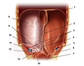
Anatomy - Ramirez component separation technique - general and visceral surgery
You have not purchased a license - paywall is active: to the product selection
Anatomy of the anterior abdominal wall
1. Layered anatomy
The large area of the anterior abdominal wall from the xiphoid process and both costal arches to the pelvis demonstrates a characteristic layered anatomy: Skin covering the subcutaneous fatty tissue; superficial fascias; muscles and their fascias; and then an extraperitoneal fascia and the parietal peritoneum.
Particularly in the anterior wall inferior to the umbilicus the otherwise single-layered superficial fascia transitions into a double-layered structure (panniculus adiposus abdominis) comprising a superficial fatty layer (Camper fascia) and a deeper membranous layer (Scarpa fascia). The five abdominal muscles comprise:
- Three oblique muscles (1. external oblique, 2. internal oblique and 3. transversus abdominis) Two straight muscles (4.
- rectus abdominis and the inconsistent 5. pyramidalis muscle).
a) Superficial muscle
It courses directly posterior to the superficial fascia in inferomedial direction to the large aponeurosis; both sides conjoining in the middle to the linea alba (white line). Its inferior margin constitutes the inguinal ligament from the anterior superior iliac spine to the pubic tubercle; medially, the inguinal ligament in turn gives off the lacunar and pectineal ligament (Cooper) respectively.
b) Middle muscle
The fibers course superomedially and conjoin with the fibers from 1. to the linea alba.
c) Deepest muscle
Transverse fibers, also conjoining with the linea alba.
- Both the anterior and posterior aspect of each of the three oblique abdominal muscles is covered by its own thin fascia, and 3. at its posterior aspect the strong transversalis fascia. It invests the abdominal cavity and cephalad becomes the diaphragmatic fascia and posteriorly the thoracolumbar fascia. Caudad it attaches to the iliac crest and becomes the endopelvic fascia.
- Long, straight, paired abdominal muscle divided by 3 – 4 transverse tendinous intersections ("six-pack")
- Triangular rudimentary muscle caudad and anterior to 4. between the pubic bone and linea alba.
2. Fascias and peritoneum
Muscles 4. and 5. are invested by the rectus sheath formed by the 3 oblique abdominal muscles 1.–3. The superior ¾ of 4. are completely invested by the rectus sheath, while the latter only covers the anterior aspect of the inferior ¼ below the arcuate line. The posterior aspect of 4. is only covered by the transversalis fascia and peritoneum. In its superior ¾ the anterior lamina of the rectus sheath is formed by 1. and half of 2., while the posterior lamina is formed by both 2. and 3.
Transversalis fascia and peritoneum are separated by the extraperitoneal space, the extent of which may markedly differ depending on the location. The retroperitoneal abdominal organs are found in the retroperitoneum. At the anterior abdominal wall the slender retroperitoneum often is known as the preperitoneal space (e.g., at the deep inguinal orifice).
The peritoneum (parietal serosa) invests the completely enclosed peritoneal cavity (exception: tube openings in women), and as visceral peritoneum it invests the intraperitoneal organs. Inferior to the umbilicus it forms three folds:
- The single median umbilical fold (obliterated urachus/allantoic stalk)
- The paired medial umbilical fold (former umbilical artery)
- The paired lateral umbilical fold (inferior epigastric vessels)
3. Innervation and blood supply
The anterior abdominal wall is innervated by the anterior branches of spinal nerves Th7 – Th11, Th12 (subcostal nerve) and L1 (iliohypogastric nerve and ilioinguinal nerve).
Its cephalic superficial blood supply comes from the internal thoracic artery (→ musculophrenic artery), while caudad the blood is supplied by the superficial epigastric artery and superficial circumflex iliac artery (both branches of the femoral artery). The deep layers are supplied cephalad by the superior epigastric artery (internal thoracic artery), laterally from the intercostal vessels and caudad from the inferior epigastric artery and deep circumflex iliac artery (both branches of the external iliac artery). Venous drainage is via the corresponding (eponymous) veins.
Single Access
Access to this lecture
for 3 days
€4.99 inclusive VAT
webop-Account Single
full access to all lectures
price per month
for the modul: vascular surgery