Transabdominal preperitoneal patch (TAPP) repair of inguinal hernia - general and visceral surgery
You have not purchased a license - paywall is active: to the product selection
Principle
Accomplishing the pneumoperitoneum
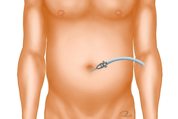
Trocar positions
After access to the abdominal cavity with the scissors, bluntly insert the trocar (10 mm) for the camera into the abdominal cavity and perform the initial inspection. At about the lateral level of the umbilicus insert a 5 mm trocar on the contralateral side of the hernia, while the ipsilateral side requires either a 5 mm or 10 mm trocar.
Tip: The 30°/45° laparoscope with the camera should face anteriorly. This allows the surgeon to survey the posterior wall of the inguinal canal.
Reducing the contents of the hernia sac
Incising and dissecting the peritoneum, exposing the anatomical landmarks
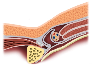
Transect the peritoneum in curved fashion superior to the hernia orifice from the medial umbilical fold to several centimeters lateral to the deep inguinal ring. Medial to the epigastric vessels, dissect between the bladder (Caution: Bladder injury!) and the posterior wall of the rectum up to the pectineal ligament (Cooper) and expose the symphysis. Now bluntly free the peritoneum caudad off the epigastric vessels, spermatic duct and the vessels of the spermatic cord.
Tip: Start the incision well superior to the deep inguinal ring or well away from the hernia sac respectively.
Exposing and reducing the contents of the hernia sac, preparing the mesh support
Completely expose and reduce the hernia sac sparing the vessels (spermatic cord, epigastric, corona mortis).
Identify any structures to be used for mesh support later on and all hernia orifices.
Tip: Dissect the peritoneum off the vessels and spermatic duct over a sufficient distance since otherwise the mesh might flip up and over.
Mesh insertion
Mesh fixation
Avoiding any tension and with just a few staples, fixate the mesh to the pectineal ligament (Cooper) and the superior medial region of the anterior abdominal wall.
As demonstrated in this example, another option would be to fixate the mesh with tissue sealant.
When stapling beware of the following:
Postoperative neuralgia may be avoided by inserting the staples neither inferior to the inguinal ligament nor lateral to the epigastric vessels. To rule out vascular injury (corona mortis), fixation at the pectineal ligament (Cooper) requires a clear view.
Spare the following nerves which always course lateral to the epigastric vessels: lateral femoral cutaneous, ilioinguinal, iliohypogastric, genitofemoral (femoral and genital branches)!
Depending on the toxicity of any tissue sealant used beware where the above nerves course.
Note: For synthetic meshes synthetic tissue sealants may be used. The biomesh in the present example requires a fibrin tissue sealant.
Closing the peritoneal incision
Lower the pressure of the pneumoperitoneum to 6-8 mmHg and close the peritoneal incision with a running V-Loc suture 3/0.
Remove the trocars under visual control and let the pneumoperitoneum escape. Suture the fascia at each incision > 5 mm from the outside, with percutaneous preplaced fascia sutures (suture catcher) if needed.
Tip: When inserting a Redon drain down to the mesh, this is accomplished through the ipsilateral trocar incision and a small peritoneal incision; secure the drain with a skin suture.
webop-Account Single
full access to all lectures
price per month
for the modul: vascular surgery