Anatomy - Lichtenstein Repair of Inguinal Hernia

  1. Anatomy of the inguinal region

    Anatomy of the inguinal region

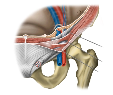
    The inguinal region (where the anterior abdominal wall transitions to the lower extremity) possesses several weak spots where a hernia sac (with or without contents) may protrude through the abdominal wall (femoral hernias are more frequent in women, while inguinal hernias are more common in men). Inferior to the inguinal ligament the septum of the iliopectineal arch divides the inguinal canal into a vascular and muscular compartment – lacuna vasorum and lacuna musculorum respectively.

    Vascular compartment

    • Both the external iliac artery and vein (→ femoral artery and vein; with the artery lateral to the vein) course through the vascular compartment which is lateral to the pubic bone. In addition, the femoral branch of the genitofemoral nerve passes through the very lateral part of this compartment, while the deep inguinal lymph nodes (Rosenmueller nodes) are located inferomedially. In femoral hernias the lacuna vasorum is the deep weak spot (through the femoral septum along the femoral vein).

    Muscular compartment

    • Lateral to the lacuna vasorum, the muscular compartment is traversed by the psoas major and iliacus muscles (together they comprise the iliopsoas muscle), the femoral nerve, and lateral femoral cutaneous nerve.
  2. Anterior abdominal wall and inguinal canal

    Anterior abdominal wall and inguinal canal

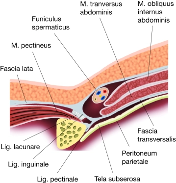
    During testicular development the tubular structure of the inguinal canal is formed by the externalization of the testicles, which are pulled into the scrotum by the gubernaculum testis. The peritoneum pulled along during this descent remains in the inguinal canal as a pouch (processus vaginalis testis). Thus, the layers of the abdominal wall turn into the corresponding sheaths in this pouch:

    Transversalis fascia → internal spermatic fascia,

    Internal oblique → cremaster,

    Fascia of external oblique → external spermatic fascia,

    No investment by the transversus abdominis since it terminates more cephalad.

    The blood vessels (testicular artery and vein), spermatic duct (ductus deferens) and nerves (ilioinguinal nerve originating from the lumbar plexus) are also pulled into the scrotum, and together they form the spermatic cord. When the processus vaginalis testis closes, it obliterates the connection with the peritoneal cavity, typically leaving behind only the vestige of the vaginal process. In the female embryo the pull of the gubernaculum does not result in a complete descent of the ovaries, which remain close to the uterus, but rather the remains of the original gubernaculum persist in the inguinal canal as the round ligament of uterus. Incomplete obliteration of the processus vaginalis testis is a weak spot in the abdominal wall and therefore a possible starting point for inguinal hernia.

    The approximately 4 cm long oblique inguinal canal, slanting downwards and medially, parallels the inguinal ligament immediately superior to it, with the cephalic end of the canal originating at the deep inguinal ring and the opening to the external abdominal wall at the superficial inguinal ring.

    Deep inguinal ring

    • Halfway between the pubic symphysis and the anterior superior iliac spine in the inner abdominal wall, lateral to the inferior epigastric artery and vein (within the lateral umbilical fold)

    Superficial inguinal ring

    • Superior to the pubic tubercle in the fascia of the external oblique, superior edge pointing cephalad, lower edge formed by inguinal ligament, sides = medial and lateral crus with stabilizing intercrural fibers.
  3. Walls of the inguinal canal

    Anterior wall

    • Fascia of the external oblique, with lateral augmentation by fibers of the internal oblique → inguinal ligament and → cremaster

    Posterior wall

    • Transversalis fascia, with medial augmentation by the conjoint tendon (=  tendon of the transversus abdominis and internal oblique).

    Superior wall

    • Transversus abdominis and internal oblique (fibers running from inguinal ligament to the medial conjoint tendon)

    Inferior wall

    • Medial inguinal ligament (= reflected ligament) and a trough for the spermatic cord formed by the external oblique.
  4. Contents of the inguinal canal

    In men: Spermatic cord (funiculus spermaticus) with the spermatic duct; deferential artery (branch of the inferior vesical artery) and testicular artery (from the aorta); venous pampiniform plexus; cremasteric artery and vein; genital branch of the genitofemoral nerve to the cremaster muscle; sympathetic nerve fibers; and lymphatic vessels. All invested by the internal spermatic fascia, cremasteric fascia, and external spermatic fascia.

    In women: Round ligament of uterus passing from the uterus through the deep inguinal ring into the inguinal canal and then on through the superficial inguinal canal to the labia majora; lymphatic vessels; and in both sexes sometimes the ilioinguinal nerve.

  5. Types of hernias

    The deep inguinal ring is the weak spot for indirect hernias, while direct hernias originate in the middle inguinal fossa (medial to the deep inguinal ring and the inferior epigastric artery and vein).

    Indirect inguinal hernia

    • More common; men > women; congenital (patent processus vaginalis) or acquired (also via the deep inguinal ring in the lateral inguinal fossa, mostly in adults); hernia sac passes lateral to the epigastric vessels into the scrotum and labia majora respectively

    Direct inguinal hernia

    • Mostly acquired; in adults men > women; pouching of the peritoneum and transversalis fascia in the middle inguinal fossa (inguinal triangle, medial to the epigastric artery and vein); delimited medially by the transversus abdominis and inferiorly by the inguinal ligament; emerges most often through the superficial inguinal ring → scrotum/ labia majora.

    Femoral hernia

    Mostly acquired; in adults women > men; together with the femoral artery and vein through the femoral canal in the medial thigh (medial to the lacuna vasorum); femoral branch of the genitofemoral nerve; and lymphatic vessels.