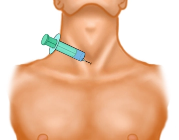
Are you sure you want to perform this action?
Percutaneous dilatational tracheotomy
You have not purchased a license - paywall is active: to the product selection
Single Access
Access to this lecture
for 3 days
€4.99 inclusive VAT

webop-Account Single
full access to all lectures
price per month
for the modul: vascular surgery
from 8,17 €
-
Anterior triangle of the neck
Situated between the anterior margin of the sternocleidomastoid muscle, the mandibula und the jugular fossa, the anterior cervical triangle near the hyoid bone comprises the suprahyoid and subhyoid muscles, vessels, nerves and the thyroid.
Fascial layers
The skin of the anterior triangle of the neck covers several fascial layers (all belonging to the cervical fascia) with distinctive features:
- The superficial lamina invests all structures of the neck, except for the platysma, and separately invests the sternocleidomastoid muscle as well as the posterior aspect of the trapezius muscle (accessory nerve XI),
- with the medial pretracheal lamina investing the subhyoid muscles and
- the deep prevertebral lamina coursing outside the surgical field between the esophagus and spine.
Just like the lateral vascular and nerve pedicle (carotid artery, internal jugular vein and vagus nerve), the trachea and thyroid / parathyroids also have their own organ fascias. With their three-dimensional configuration, the fascias invest compartments interspersed with spaces which extend into the mediastinum and thus represent potential routes of infection.
Single Access
Access to this lecture
for 3 days
€4.99 inclusive VAT

webop-Account Single
full access to all lectures
price per month
for the modul: vascular surgery
from 8,17 €
Single Access
Access to this lecture
for 3 days
€4.99 inclusive VAT

webop-Account Single
full access to all lectures
price per month
for the modul: vascular surgery
from 8,17 €
-
Local anesthesia of the surgical field
-
Bronchoscopic overview
-
Tracheal puncture and introduction of the Seldinger guidewire
Puncture in a strictly median and slightly caudad direction.
Concurrent bronchoscopic monitoring helps prevent puncture of the posterior tracheal wall or the cuff of the endotracheal tube and verify the correct position of the puncturing needle.
When air is aspirated into the syringe, the needle is within the tracheal lumen.
-
Skin incision and insertion of the dilator
-
Dilating the pretracheal tissue
-
Dilating the trachea under bronchoscopic monitoring
-
Inserting and fixating the tracheotomy tube
-
Bronchoscopic verification of tube position
As final step bronchoscopically verify the tube position, rule out tracheal injury and suction any blood and secretions.
Note: While the cuff is deflated, the bronchoscope may be advanced through the larynx along the outside of the tracheotomy tube until it reaches the tracheal bifurcation; during retraction, the entire length of the posterior membranous wall can be inspected.
Single Access
Access to this lecture
for 3 days
€4.99 inclusive VAT

webop-Account Single
full access to all lectures
price per month
for the modul: vascular surgery
from 8,17 €
Single Access
Access to this lecture
for 3 days
€4.99 inclusive VAT

webop-Account Single
full access to all lectures
price per month
for the modul: vascular surgery
from 8,17 €
-
Literature summary
-
Textbooks and metaanalyses
-
References on this topic
-
literature search
Literature search under: http://www.pubmed.com