Cephalic pancreatoduodenectomy with Blumgart anastomosis and biliopancreatic separation (Merheim technique) - general and visceral surgery
You have not purchased a license - paywall is active: to the product selection
Laparotomy
Surgical access is gained via a standard transverse incision. Follow this with inspection and palpation of the entire abdomen to rule out distant metastases and signs of local unresectability.
After inserting a wound edge protector mount the cable winch retractor system.
Note:
In patients with a sharply angled costal arch perform a midline laparotomy.
Opening the lesser sac
Enter the omental bursa by detaching the omentum from the right colic flexure to the left third of the transverse colon. Now that the pancreas can be examined in detail from the left too, look for any infiltration of the stomach or postpyloric duodenum. Take down any adhesions between the pancreas and posterior gastric wall.
Note:
The duodenum should be preserved over a distance of 2–3 cm. If this is not possible, perform partial gastric resection.
Kocher maneuver
First, detach the right colic flexure from the anterior aspect of the duodenum and pancreatic head. Then mobilize the duodenum with the Kocher maneuver. Transpose the mobilized colon section to the left, and after incising along the outer margin of the duodenum release the duodenum together with the head of the pancreas from its retroperitoneal attachments. Continue mobilizing the head of the pancreas across the aortic plane up to the ligament of Treitz until the pancreatic head can be completely enclosed and palpated from posterior.
Next, palpate the mobilized pancreatic head to rule out retroperitoneal infiltration and also palpate the lymphatic pathways and large vessels.
Cholecystectomy
Start the resection phase with cholecystectomy: Grasp the gallbladder and perform antegrade subserous dissection from the liver bed using bipolar forceps, expose the bile duct and cystic artery, which for now are left attached to the specimen later resected.
Note:
To prevent ascending cholangitis following bilioenteric anastomosis, cholecystectomy is mandatory for functional reasons when performing pancreatic head resection.
Dissecting the hepatoduodenal ligament and transecting the right gastric artery
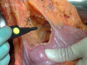
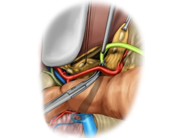
Dissection of the hepatoduodenal ligament includes exposure of the common hepatic artery, proper hepatic artery, and gastroduodenal artery, portal vein, and common bile duct.
The case presented here has an anatomical variant of the blood supply to the liver. An atypical right hepatic artery from the superior mesenteric artery crosses the common bile duct posteriorly.
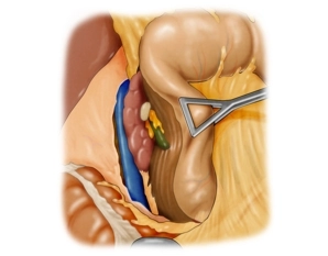
In this case, perform the lymphadenectomy along the left hepatic artery in a central direction towards the celiac trunk, exposing the bifurcation of the gastroduodenal artery.
Begin mobilizing the pylorus by dividing the right gastric artery between ligatures.
Before this, complete ligament dissection by exposing the suprapancreatic portal vein until posterior to the pancreas.
Transecting the postpyloric duodenum and gastroduodenal artery
Transecting and skeletonizing the first jejunal loop
Pulling through the jejunal limb / dissecting the superior mesenteric vein
Transecting the pancreas and CBD
Detaching the pancreatic head
Sealing the retroperitoneal resection area with HaemoCer PLUS
Oversewing and pulling up the jejunal limb retrocolically
Pancreatojejunostomy I (Blumgart-style, posterior wall)
Pancreatojejunostomy II (Blumgart-style, anterior wall)
Fashioning the Roux-en Y limb
Bilioenteric anastomosis
Anastomosing the stomach
Terminolateral jejunojejunostomy (Roux-en-Y)
Closing the abdominal wall
Single Access
Access to this lecture
for 3 days
€4.99 inclusive VAT

webop-Account Single
full access to all lectures
price per month
for the modul: vascular surgery