Karydakis flap procedure for pilonidal sinus - general and visceral surgery

You have not purchased a license - paywall is active: to the product selection
  • Planning the resection

    Video
     
    Planning the resection
     

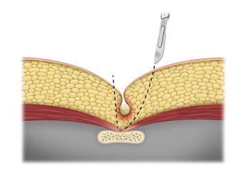
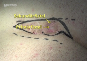
    The technique is demonstrated on a chronic recurrent pilonidal sinus.

    First, define the resection margins. The dotted lines correspond to the highest point of the buttock in question. The ovoid incision should be located eccentric to the midline and cover the entire system of tracts. The central axis of the incision should be about 2cm lateral to the midline.

  • Skin incision

    Video
     

    Incise the tissue to be removed. Since the chronic infection always predestines this operation to bleeding, we prefer the electrocautery. 

  • En-bloc excision

    Paid content (video)
    Paid content (image)

    Excise the inflamed tissue together with all the fistula tracts. On the far side of the midline bevel the incision, while bringing it straight down close to the midline. Leave a thin layer of fat on the sacral fascia.

  • Mobilizing the skin flap with fat

    Paid content (video)
    Paid content (image)

    With electrocautery or scissors mobilize the skin-fat flap from the gluteal fascia close to the midline. This allows adequate mobilization of the flaps without having to incise the fascia, thereby permitting asymmetric closure.

    After diligent hemostasis, place a Redon drain into the wound cavity.

  • Wound closure

    Paid content (video)
    Paid content (image)

    After loosening/cutting the medical tape adhesive strips on the buttocks, close the wound in layered fashion.

    With strong full-thickness three-point sutures and including the right sacral fascia, join the mobilized skin-fat flap with the contralateral wound margin. This will largely prevent cavity formation.

    Tip: It is best to first place the stitches without tying them.

    Follow this with a subcutaneous suture and close the skin with interrupted sutures.

    And as the final step, instill some local anesthetic through the Redon drain.

    Note: Suture line and later scar are about 1-2cm off the midline. The natal cleft has flattened out somewhat, particularly in its superior region, making it more difficult for hair to penetrate the skin once more.

date of publication: 27.03.2013

webop-Account Single

full access to all lectures
price per month

for the modul: vascular surgery

from 8,17 €

hospitals & libraries

for the modul: vascular surgery

from 390,00 euros