The large area covering the anterior abdominal wall from the xiphoid process or the costal arches to the pelvic bones has a typical layered structure: beneath the skin and subcutaneous fat tissue, there are superficial fasciae, muscles and their fasciae, then an extraperitoneal fascia and the parietal peritoneum.
Especially in the anterior wall below the navel, the otherwise typical single-layered superficial fascia transitions into a two-layered structure (Panniculus adiposus abdominis) consisting of a superficial fat-rich layer (Camper's fascia) and a deeper membranous layer (Scarpa's fascia). The 5 abdominal muscles consist of:
3 oblique muscles (1. External oblique muscle, 2. Internal oblique muscle, and 3. Transverse abdominal muscle)
2 straight muscles (4. Rectus abdominis muscle and the inconstant 5. Pyramidalis muscle).
- external muscle
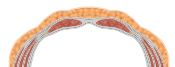
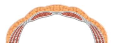
located immediately beneath the superficial fascia, it runs caudomedially to the large aponeurosis, both sides merge centrally to form the linea alba. Its lower edge forms the inguinal ligament from the anterior superior iliac spine to the pubic tubercle, which in turn medially branches into the lacunar ligament and the pectineal ligament (Cooper's ligament). - middle muscle
fibers run craniomedially merging with fibers from 1. to the linea alba. - innermost muscle
transversely running fibers, also radiating into the linea alba.
Each of the three oblique abdominal muscles has a thin, own fascia on its front and back surface, 3. on its inner side the strong transversalis fascia. This lines the abdominal cavity and transitions cranially into the diaphragmatic fascia and dorsally into the thoracolumbar fascia. Caudally it is attached to the iliac crest and transitions into the endopelvic fascia.
Long straight paired abdominal muscle, interrupted by 3 – 4 transverse tendinous intersections ("six-pack").
Triangular rudimentary muscle caudal and ventral to 4. between the pubic bone and linea alba.