Are you sure you want to perform this action?
Esophageal resection
You have not purchased a license - paywall is active: to the product selection
Single Access
Access to this lecture
for 3 days
€4.99 inclusive VAT

webop-Account Single
full access to all lectures
price per month
for the modul: vascular surgery
from 8,17 €
-
Surgical anatomy of the esophagus/stomach
In terms of function, the stomach mixes and stores food and is an expansion of the alimentary tract between the esophagus and the duodenum. This muscular hollow viscus produces acidic gastric juice (mucus and HCl) and enzymes, which predigest some elements of the ingested food, and portions the chyme into the duodenum.
Usually, the stomach is located immediately inferior to the diaphragm in the left upper quadrant and epigastrium. Location, size and shape of the stomach vary from person to person and may differ substantially, depending on age, filling condition and body position. The moderately filled stomach has a mean length of 25-30 cm and can hold 1.5 liters, in extreme cases up to 2,5 liters.
Within the abdominal cavity the stomach is held in position and stabilized by ligaments inserting at the liver and spleen Its convex aspect forms the major curvature (curvatura major gastrica) and its concave aspect the lesser curvature (curvatura minor gastrica). Its anterior wall is termed paries anterior gastrica and its posterior aspect paries posterior gastrica.
Since the stomach is an intraperitoneal viscus, it is covered by the gastric serosa (tunica serosa gastrica), and only the posterior aspect of the cardia is free of serosa. Stomach rotation shifts the embryonic mesogastrics from their former sagittal position to a frontal location. The lesser omentum originates at the lesser curvature and extends to the hepatic portal, while the greater omentum originates at the greater curvature and courses to the transverse colon, spleen and diaphragm.
The stomach displays the following portions:
- Entrance of stomach / Cardia / Ostium cardiacum:
The superior opening of the stomach, where the esophagus enters the stomach, is 1-2 cm long. It is characterized by a marked transition from the mucosa of the esophagus to that of the stomach. - Gastric fundus / fundus gastricus:
Superior to the level of entrance of the esophagus the fundus arches cephalad, which then is called gastric fornix (fornix gastricus). Usually, the fundus is full of air which is swallowed automatically when ingesting food. In the erect position the fundus is the highest point of the stomach, and on abdominal films its trapped air is evident as the “gastric bubble”. A notch (incisura cardialis) clearly delimits the fundus from the entrance of the stomach. - Body of the stomach / Corpus gastricum
The main portion of the stomach is taken up by the gastric body. The deep mucosal folds (plicae gastricae) found here extend from the cardia to the pylorus and are also known as “magenstrasse”. - Pylorus / Pars pylorica:
This portion begins with the extended pyloric antrum, followed by the pyloric canal, and terminates at the actual pylorus. It is formed by the pyloric sphincter (m. sphincter pyloricus), a strong circular layer of muscle which closes off the inferior gastric orifice (ostium pyloricum). The pylorus closes off the gastric outlet and periodically lets some of the chyme pass into the adjacent duodenum.
- Entrance of stomach / Cardia / Ostium cardiacum:
-
Function
The stomach acts as a reservoir for ingested food. Since it may store food for hours, it ensures that we can meet our daily nutritional requirements with a few major meals. Peristalsis thoroughly mixes the chyme with the gastric juice, the food is broken up chemically, predigested and then portioned into the duodenum.
-
Duodenum
The first of four portions of the duodenum, the bulb, begins at the pylorus and transitions into the second portion, the descending part. The horizontal portion begins at the inferior duodenal flexure and sweeps to the left. The ascending portion transitions into the jejunum at the duodenojejunal flexure (ligament of Treitz) The blood supply to the duodenum is provided by the gastroduodenal, pancreaticoduodenal and supraduodenal arteries, which originate from the celiac axis and the superior mesenteric artery.
Single Access
Access to this lecture
for 3 days
€4.99 inclusive VAT

webop-Account Single
full access to all lectures
price per month
for the modul: vascular surgery
from 8,17 €
Single Access
Access to this lecture
for 3 days
€4.99 inclusive VAT

webop-Account Single
full access to all lectures
price per month
for the modul: vascular surgery
from 8,17 €
-
Laparotomy and exploration
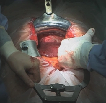
After checking patient position, skin prepping and sterile draping make a midline skin incision from the xiphoid to the navel, passing the latter on the left. After transecting the subcutaneous tissue with electrocautery open the abdominal cavity along the linea alba.
Insert a circular wound edge protector, thoracic retractor and the Ulrich cable winch retractor.
Explore the entire abdominal cavity to rule out peritoneal and liver metastasis, if necessary, by ultrasonography.
-
Mobilizing the intraabdominal esophagus
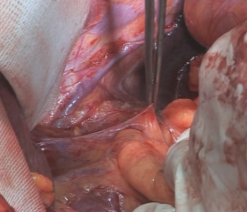
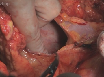
Incise the lesser omentum close to the liver. As shown here, spare any atypical left hepatic artery arising from the left gastric artery. Continue the incision onto the pre-esophageal peritoneum and bluntly open the esophageal hiatus within the tissue layer separating the right crus of diaphragm from the esophagus. Expose the esophagus craniad and to the left crus of diaphragm by blunt dissection. Loop the esophagus with a Penrose drain or like to put traction on it.
-
Opening the esophageal hiatus and dissecting the distal esophagus
-
Freeing the duodenum
In order to achieve a tension-free gastric transposition, perform a generous Kocher maneuver to free the duodenum. Starting laterad, perform this maneuver until the vena cava and the medial aspect of the aorta are exposed. Now both the duodenum and head of the pancreas are mobile enough that the pylorus can be guided craniad into the esophageal hiatus without difficulty.
-
Opening the omental bursa
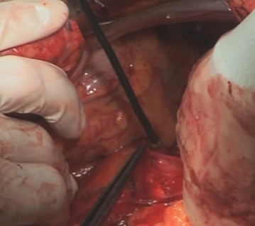
Free the greater omentum from the transverse colon and open the lesser sac. Initially leave the greater omentum along the greater curvature. Divide the greater omentum at the gastrolienal ligament near the spleen between Overholt forceps.
Tip:
- During cranial dissection, pack an abdominal towel behind the spleen to prevent any traction on it, which otherwise might tear the spleen. Whenever the situation is ambiguous, be generous with the indication for splenectomy.
-
Freeing the stomach
-
Abdominal lymphadenectomy
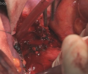
Best start the suprapancreatic lymphadenectomy (compartment II) at the celiac trunk by exposing the gastroduodenal artery. Follow it up to its junction with the common hepatic artery. Take down all covering lymph nodes and lymph vessels along the common hepatic artery.
Tip:
- Dissection of the hepatoduodenal ligament is simplified by passing the left index finger through the omental foramen (Winslowii).
Continue the lymphadenectomy along the splenic artery into the left upper quadrant. Since there is direct drainage via the retroperitoneum to the hilum of the left kidney, we recommend continuing the dissection up to the renal vein.
-
Dividing V. gastrica dextra/sinistra and left gastric artery
When dissecting the common hepatic artery mediad V. gastrica dextra/sinistra is encountered, which is divided between clips/ligatures.
In the presence of an atypical left hepatic artery arising from the left gastric artery, the latter cannot be suture ligated and divided between Overholt forceps close to its origin, but rather after the left hepatic artery has branched off.
-
Dividing the greater omentum
Divide the greater omentum along the greater curvature outside the arterial and venous gastroepiploic arcades. In doing so, the right gastroepiploic artery and vein, which parallel the greater curvature in a fatty envelope from the inferior aspect of the pylorus toward the gastric body, must be preserved at all costs.
When taking down the greater omentum step by step toward the gastric fundus, this is facilitated by grasping the greater omentum and lifting it out of the incision distally. Care must be taken to preserve the rather delicate arcade between the left and right gastroepiploic arteries to prevent any impairment of the blood supply at the tip of the gastric tube constructed later.
-
Constructing the gastric tube
The blood supply of the stomach via the right gastroepiploic artery is rather varied but should suffice for the blood supply of the gastric tube to be constructed. It nevertheless is advisable to preserve the right gastric artery arising from the hepatic artery proper in order to ensure adequate blood supply of the gastric tube.
Start constructing the gastric tube along the lesser curvature where the Latarjet nerve (the so-called crow's foot of the vagus nerve) enters the gastric wall.
Divide the stomach by multiple applications of the linear stapler (GIA 60).
Tip:
- Overlap the individual staple lines somewhat to prevent necrosis and gaps.
In terms of postoperative function, it is important to keep the lumen of the gastric tube as small as possible, with a maximum patency of 3-4 cm.
-
Suturing and transposing the gastric tube
-
Thoracotomy
-
En-bloc esophagectomy
-
Gastric transposition and intrathoracic anastomosis
-
Enveloping the anastomosis and shortening the gastric tube
-
Closing the thoracotomy
-
Placing a feeding tube
-
Closing the abdominal wall
Single Access
Access to this lecture
for 3 days
€4.99 inclusive VAT

webop-Account Single
full access to all lectures
price per month
for the modul: vascular surgery
from 8,17 €
Single Access
Access to this lecture
for 3 days
€4.99 inclusive VAT

webop-Account Single
full access to all lectures
price per month
for the modul: vascular surgery
from 8,17 €
-
Literature summary
-
Ongoing trials on this topic
-
References on this topic
-
Reviews
-
Guidelines
-
literature search
Literature search under: http://www.pubmed.com