Total laparoscopic gastrectomy with D2 lymphadenectomy - general and visceral surgery
You have not purchased a license - paywall is active: to the product selection
Trocar positioning
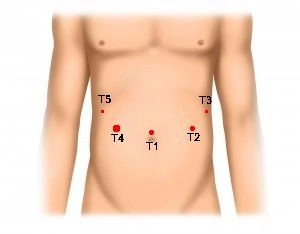
Start the procedure by marking the trocar positions on the abdominal wall. After inserting the Veress needle and pressure-controlled CO2 insufflation, place the following trocars in a semicircle:
- T1 = camera trocar (10 mm)
- T2 = working trocar(10 mm)
- T3 = working trocar 5 mm (later 12 mm for CEEA stapler and extension of incision for specimen retrieval)
- T4 = working trocar (12 mm)
- T5 = working trocar (5 mm)
Incising the gastrocolic ligament and resecting the omentum (LN 4)
Dissecting the duodenum (LN 11) and transecting the right gastric artery
Continue the dissection at the gastroduodenal junction. Mobilize the duodenum likewise with the Thunderbeat® device to around 2 cm distal to the pylorus. After lifting the stomach, locate the right gastric artery and divide it close to its origin at the proper hepatic artery.
Note: Pay particular attention when dissecting the posterior aspect of the duodenum. Make every attempt not to injure the pancreatic capsule as this could result in pancreatic fistula, local inflammatory reaction, and leakage from the duodenal remnant.
Dissecting the common hepatic artery, resecting the lesser omentum and transecting the duodenum
Exposing the celiac trunk (LN 8), dissecting and dividing the left gastric artery
Dissecting the splenic hilum (LN 10)
Dissecting the esophageal hiatus, including lymphadenectomy down to the aorta (LN 1 and 2)
Temporarily anchoring the jejunum on the stomach, Roux-en-Y jejunostomy
Restore passage with the esophagus in end-to-side technique with the circular EEA stapler by introducing it into the open end of the Roux limb (“shepherd’s crook”). Prepare the jejunojejunostomy by locating a jejunal limb about 30 cm distad of the duodenojejunal flexure (ligament of Treitz), pull it up loosely to the stomach anterior to the colon, and temporarily anchor it with a suture at the gastric fundus. Follow the efferent limb of this anchored loop for another 50–60 cm and then position it such that both limbs parallel each other. After punctate antimesenteric opening of both intestinal lumens with the electrocautery hook, fashion the side-to-side anastomosis with a 60 mm Endo-GIA stapler. Close the insertion site of the stapler halves with a running suture Vicryl 3/0.
Note: The reconstruction presented here is an established surgical technique used in bariatric laparoscopic gastric bypass.
Opening the esophagus and placing the CEEA anvil through the mouth
First, insert via the mouth a nasogastric tube mounted on the anvil of a 21 mm CEEA stapler (nasogastric tubes with pre-mounted anvils are available as ready-made medical products in sizes 21–25 mm from the medical industry). Next, open the esophagus by semicircular transection of its anterior wall just above the cardia. Once the tip of the gastric tube appears in the incised esophagus, pull it through into the abdominal cavity Now arm the semicircular opening in the esophagus with a monofilament purse string suture (nonabsorbable, size 0). Next, complete the esophageal transection, pull through the anvil into the distal esophagus and secure it with the purse string suture. Finally, detach the nasogastric tube from the anvil and withdraw it from the abdominal cavity.
End-to-side esophagojejunostomy
Remove the suture anchoring the jejunum at the gastric fundus. Transect the pulled-up first jejunal limb 10–15 cm distad of the side-to-side jejunojejunostomy. After extending the trocar incision in the left upper quadrant (T3), insert the 21 mm CEEA stapler first into the abdominal cavity and then into the opened intestinal lumen. Pull up the limb to the esophagus anterior to the colon, perforate the intestinal wall at the antimesenteric border with the spiked shank and then insert the extended spike into the anvil shaft. When closing the stapler, it became evident that the esophageal wall was not properly centered due to inadequate tension. Therefore, this had to be corrected by a second purse string suture (V-Loc 2/0) which was tightened step by step. Problem-free mechanical fashioning of the esophagojejunostomy was now possible.
Completing the Roux-en-Y anastomosis with jejunal segment resection
Oversewing esophagojejunostomy suture line failure
Retrieving the resected gastric specimen, drainage
Resected gastric specimen
webop-Account Single
full access to all lectures
price per month
for the modul: vascular surgery