Esophageal cancer is one of the leading causes of cancer-related deaths worldwide. Recent advances have led to multidisciplinary treatment approaches, with a particular focus on neoadjuvant therapies. Nevertheless, surgical therapy remains the primary treatment option with a curative approach. Despite advances in surgical technique, esophageal surgery is associated with a high risk of morbidity and mortality, leading to prolonged hospital stays and increased healthcare costs. One of the most severe postoperative complications is anastomotic leakage, with an incidence of over 10%.
A similarly challenging scenario arises with any transmural defect of the esophagus. Esophageal leaks lead to life-threatening complications such as mediastinitis, pleural empyema, sepsis, or bronchial erosion.
The treatment strategy for defects of the upper gastrointestinal tract is not standardized. Over the years, the approach has shifted from surgical intervention to endoscopic interventions.
Surgical measures are generally limited to emergencies with septic unstable patients and/or necrosis of the gastric conduit, with high morbidity and mortality due to the invasiveness and often critical condition of the patients.
In all other cases, an endoscopic approach is preferred. Numerous endoscopic techniques have been developed, driven by growing scientific interest and technological advances in this field.
The introduction of self-expanding, fully covered metal stents (SEMS) led to a shift in treatment towards endoluminal endoscopic therapy. However, clinical success was limited, as migration and dislocation occurred in over 50% of cases, and there was no possibility for drainage.
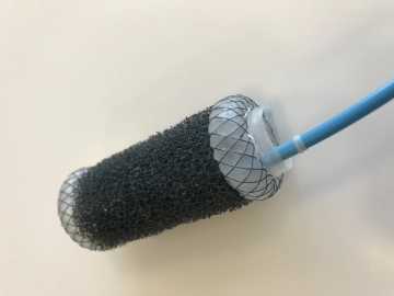
The principle of endoscopic vacuum therapy (EVT) represented a significant milestone, as it transferred the positive results of negative pressure wound therapy in secondary wound healing to the internal area of the body or the gastrointestinal tract.
The vacuum stent therapy (VAC-Stent) represents the latest technological innovation in the endoscopic management of anastomotic leaks and esophageal perforations. This device combines the advantages of both methods in a single system: It seals the leak and maintains esophageal passage for oral intake (SEMS), while simultaneously providing a drainage system for wound cavities associated with anastomotic leaks and esophageal perforations (EVT).