
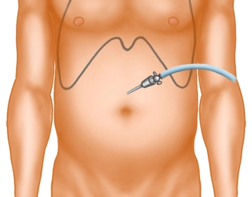
A small skin incision is made well above the navel in the midline. After inserting the Veress needle and verifying the correct position, the pneumoperitoneum is established.
-
Skin incision
![Skin incision]()
Soundsettings The video clip can be played back with the automatic soundtrack of the subtitles.
In the sidebar registered users can enable and disable the automatic start of the dubbing.
Remark: Since this is a text-to-speech computer voice, it may mispronounce some medical terminology. -
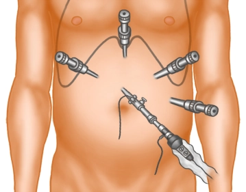
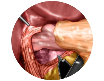
Trocar positioning
![Trocar positioning]()
Soundsettings The video clip can be played back with the automatic soundtrack of the subtitles.
In the sidebar registered users can enable and disable the automatic start of the dubbing.
Remark: Since this is a text-to-speech computer voice, it may mispronounce some medical terminology.The optics are introduced via a 5mm/10mm trocar. A diagnostic laparoscopy is performed. Under direct vision, 4 trocars are placed in the upper abdomen.
-
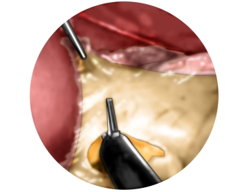
Traction of the stomach and incision of the lesser omentum
![Traction of the stomach and incision of the lesser omentum]()
Soundsettings The video clip can be played back with the automatic soundtrack of the subtitles.
In the sidebar registered users can enable and disable the automatic start of the dubbing.
Remark: Since this is a text-to-speech computer voice, it may mispronounce some medical terminology.The left liver lobe is held up with the laparoscopic probe and the stomach is tensed with the Babcock clamp. In the area of the pars flaccida, the lesser omentum is incised with the ultrasonic scalpel.
-
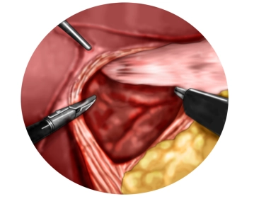
Preparation of the diaphragmatic crura with entry into the mediastinum
![Preparation of the diaphragmatic crura with entry into the mediastinum]()
Soundsettings The video clip can be played back with the automatic soundtrack of the subtitles.
In the sidebar registered users can enable and disable the automatic start of the dubbing.
Remark: Since this is a text-to-speech computer voice, it may mispronounce some medical terminology.Now the right diaphragmatic crus is displayed. Then, proceed anteriorly over the anterior commissure to the left diaphragmatic crus and display it as well. In doing so, the ventral mediastinum is opened.
-
Preparation of the lower esophagus
![Preparation of the lower esophagus]()
Soundsettings The video clip can be played back with the automatic soundtrack of the subtitles.
In the sidebar registered users can enable and disable the automatic start of the dubbing.
Remark: Since this is a text-to-speech computer voice, it may mispronounce some medical terminology.One now proceeds far into the lower mediastinum and circumferentially mobilizes the lower esophagus from its adhesions. In doing so, the posterior vagus nerve is clearly identified and remains with the esophageal musculature. The esophagus is mobilized from the mediastinum to the extent that the area of the lower esophageal sphincter is tension-free in the abdominal cavity.
Mobilization of the gastric fundus
The gastrosplenic ligament and the short gastric vessels are transected at the level of the splenic
The gastrosplenic ligament and the short gastric vessels are transected at the level of the splenic
Activate now and continue learning straight away.
Single Access
Activation of this course for 3 days.
US$9.40
inclusive VAT
Most popular offer
webop - Savings Flex
Combine our learning modules flexibly and save up to 50%.
from US$7.27 / module
US$87.34/ yearly payment
general and visceral surgery
Unlock all courses in this module.
US$14.55
/ month
US$174.70 / yearly payment