
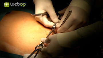
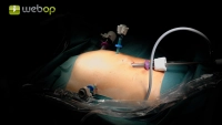
Supraumbilical mini-laparotomy, insertion of the optical trocar, and establishment of pneumoperitoneum. Under laparoscopic vision, the working trocars are placed:
5 mm left flank, 5 mm right mid-abdomen, 12 mm right lower abdomen.
Start your free 3-day trial — no credit card required, full access included
Supraumbilical mini-laparotomy, insertion of the optical trocar, and establishment of pneumoperitoneum. Under laparoscopic vision, the working trocars are placed:
5 mm left flank, 5 mm right mid-abdomen, 12 mm right lower abdomen.
The patient is placed in the Trendelenburg position so that the small intestine can be moved cranially out of the pelvis. Then, the adhesions of the sigmoid colon to the lateral pelvic wall are released.
Note: In women with an intact uterus, fixing the uterus ventrally to the abdominal wall can be advantageous.
This can be conveniently done with a straight needle. It is inserted percutaneously above the symphysis, then laparoscopically through the uterine corpus, and then back out through the abdominal wall. It is then tied extracorporeally so that the uterus is under tension.
Now the identification of the promontory takes place. At the level of the promontory, the peritoneum is opened to the right of the rectum. From here, a J-shaped incision of the peritoneum is performed. The incision is initially extended caudally pararectally to the right and then completed in an arc from right to left over the Douglas space.
Activation of this course for 3 days.
Most popular offer
Combine our learning modules flexibly and save up to 50%.
US$86.85/ yearly payment
Unlock all courses in this module.
US$173.70 / yearly payment
Webop is committed to education. That's why we offer all our content at a fair student rate.