Start your free 3-day trial — no credit card required, full access included

Gastrectomy

Reading time readingtime 24:19 min.
  1. Transverse Upper Abdominal Laparotomy

    Video
    Transverse Upper Abdominal Laparotomy
    Soundsettings

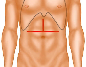
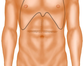
    The opening of the abdomen is performed via a transverse upper abdominal laparotomy with upper median laparotomy, so-called “inverted T“.
    Then, using diathermy, the subcutaneous tissue and subsequently the rectus musculature are transected along the linea alba.
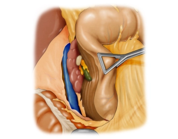
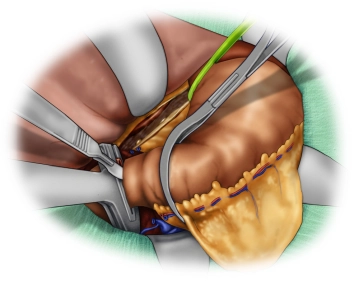
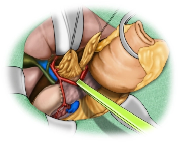
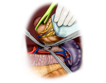
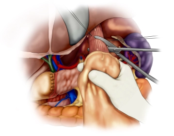
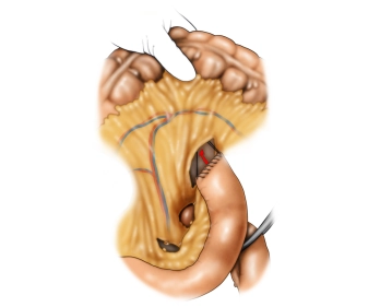
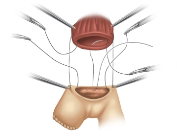
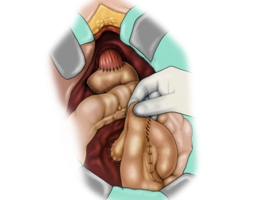
    The opened abdominal cavity is now explored to assess the location and extent of the tumor and thus the resectability after exclusion of distant metastases, especially on the peritoneum and liver. The tumor is located in the middle third of the stomach at the greater curvature.

  2. Detachment of the lesser omentum; Preparation of the gastroesophageal junction

    Video
    Detachment of the lesser omentum; Preparation of the gastroesophageal junction
    Soundsettings

    After releasing pericholecystic adhesions, the lesser omentum is detached close to the liver from distal to proximal while sparing the hepatoduodenal ligament. The preparation then continues to the gastroesophageal junction. The abdominal esophagus is circumferentially exposed and looped.

  3. Kocher Mobilization

    Video
    Kocher Mobilization
    Soundsettings

    Now follows the mobilization of the duodenum according to Kocher. In this process, the paraduodenal peritoneum is incised approximately 1 cm away from the lateral margin and the duodenum is elevated dorsally in a largely avascular plane from the retroperitoneum and the vena cava is exposed.

  4. Preparation at the hepatoduodenal ligament

    Video
    Soundsettings

    Start of the lymph node dissection at the hepatoduodenal ligament (LN group 12), during which the right gastric artery is transected close to its origin and the common hepatic artery is looped. 

  5. Detachment of the greater omentum and the gastroepiploic vessels

    Video
    Detachment of the greater omentum and the gastroepiploic vessels
    Soundsettings

    By dissecting the greater omentum from the transverse colon, the omental bursa is completely opened. In doing so, the greater omentum is left at the greater curvature. Next, the right gastroepiploic vessels are centrally divided between Overholt clamps. 

    Note: To completely mobilize the stomach, later the short gastric vessels from the gastric fundus to the spleen as well as smaller vessels on the posterior wall of the stomach and also a possibly present posterior gastric artery are divided or ligated, as well as the pars densa of the lesser omentum.

  6. Transection of the Duodenum

    Video
    Transection of the Duodenum
    Soundsettings

    Now, the proximal duodenum is circumferentially prepared and subsequently divided approximately 3 cm postpylorically using a linear stapler (not shown in the video). The staple line of the duodenal stump is secured with seromuscular interrupted sutures.

    Note: A tension-free secure closure of the duodenal stump can only be achieved if a sufficient mobilization of the duodenum according to Kocher has been performed beforehand.

  7. Lymphadenectomy along the hepatic artery

    Video
    Lymphadenectomy along the hepatic artery
    Soundsettings

    The lymphadenectomy is continued to the left along the adventitia of the retracted common hepatic artery. One first encounters the left gastric vein (formerly vena coronaria ventriculi), which is divided and ligated. Finally, one reaches the celiac trunk and can divide the left gastric artery close to its origin.

    Caution! An aberrant left hepatic artery or a larger branch of the same from the left gastric artery must be excluded beforehand.

    The lymphadenectomy is then continued para-aortally to the right up to the right diaphragmatic crus. 

  8. Lymphadenectomy along the splenic artery

    Video
    Lymphadenectomy along the splenic artery
    Soundsettings

    The lymphadenectomy is now continued and completed along the upper border of the pancreas along the splenic artery to the splenic hilum. The entire lymph node package is lifted en bloc from the upper border of the pancreas and the two arteries (common hepatic artery and splenic artery) and remains on the specimen.

    Note: The film shows the specimen still in situ at the end of the surgical step.

  9. Removal of the Specimen

    Video
    Removal of the Specimen
    Soundsettings

    After truncal vagotomy, the esophagus is openly transected and the specimen removed. The film then shows the specimen cut open and enlarged. Finally, the site after removal of the specimen is shown, with the common hepatic artery and the splenic artery still looped here.

    Note: An intraoperative frozen section examination of the esophageal resection margin is mandatory.

  10. Formation of the Roux-Y Loop

    Video
    Formation of the Roux-Y Loop
    Soundsettings

    As the next step, a jejunal loop is fashioned according to the Roux technique. To this end, the mesenteric vessels are visualized approximately 20 – 30 cm aboral to the duodenojejunal flexure under transillumination. An asymmetrical vascular pedicle is created orally, since the blood supply should primarily come from aborally. The jejunum is transected with a linear stapler (GIA) and the aboral stump is oversewn. This sufficiently long efferent jejunal loop is now transposed through a mesocolic slit into the upper abdomen.

  11. End-to-Side Esophagojejunostomy

    Video
    End-to-Side Esophagojejunostomy
    Soundsettings

    The anastomosis is created as a single-row end-to-side esophagojejunostomy by hand suture. First, the jejunal loop, which has been transposed retrocolically into the upper abdomen, is incised antimesenterically over a length of 3-5 cm. This is followed by the posterior wall sutures. All posterior wall sutures are placed first, with the knots later lying inside. These sutures are placed seromuscularly on the jejunum from inside to outside and then transmurally on the esophagus from outside to inside. Absorbable suture material of size 4/0, e.g., Vicryl, is used. The jejunum is now advanced toward the esophagus over the pre-placed posterior wall sutures with the aid of a swab on a stick. After adaptation with the posterior esophageal wall, the posterior wall sutures are tied in sequence. The anterior wall is also created in a single row using the same technique, with the knots now lying outside.

    Note:

    1. Before the suture row of the anterior wall is performed, a naso-enteral tube can be placed into the efferent jejunum (not shown in the film).

     2. Alternatively, the anastomosis can be formed with a circular stapler (EEA, 25mm).

  12. Jejunojejunostomy

    Video
    Jejunojejunostomy
    Soundsettings

    The jejunojejunostomy is planned at a distance of 40-60 cm from the esophagojejunal anastomosis. The anastomosis between the short afferent and the pulled-up Roux-Y limb is performed end-to-side. The jejunojejunostomy is performed with an absorbable suture of size 4-0 in a single-layer continuous fashion. Thus, the intestinal continuity is restored. The closure of the mesenteric defect follows (not shown in the film).

    The video shows at the end the situs at the end of the reconstruction phase. 

  13. Closure of the Abdomen

    Video
    Closure of the Abdomen
    Soundsettings

    After drainage insertion, the abdominal closure is performed using a continuous all-layer fascial suture with slowly absorbable suture material of size 0, e.g. PDS and skin closure after adequate hemostasis with single interrupted sutures.

    Note: The esophagojejunostomy is drained to the right, with the drain placed past the duodenal stump. Optionally, a second drainage can also be inserted in the area of the suprapancreatic lymphadenectomy to prevent fluid retentions due to lymph fistulas. Both are not shown in the film.

Adverts

Active local hemostasis and sealing

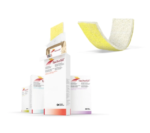
TachoSil® Versiegelungsmatrix

TachoSil® is used in adults and children from 1 month of age as supportive treatment in surgery for improving hemostasis, for supporting tissue sealing, and for suture support in vascular surgery when standard techniques are insufficient. TachoSil® is used in adults for supportive sealing of the dura mater to prevent postoperative cerebrospinal fluid leakage after neurosurgical procedures.

Produktwebsite TachoSil®
TachoSil® Prescribing Information 05-2025 (354.1 kB)

to top