| 
|
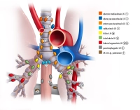
According to the IALSC lymph node map, thoracic lymph nodes are divided into 4 groups and a total of 14 stations.
- Upper mediastinal lymph nodes
Station 1 - Highest mediastinal LN - Above a horizontal line of the left brachiocephalic vein (upper edge)
Station 2 - Highest paratracheal LN - Below Station 1 and above a horizontal line at the level of the aortic arch
Station 3 - Prevascular LN - retrotracheal and prevascular LN
Station 4 - Lower paratracheal LN - between the aortic arch and the right or left main bronchus
- Aortic lymph nodes
Station 5 - Subaortic LN - Lateral to the ascending aorta and the aortic arch or brachiocephalic trunk
Station 6 - Para-aortic LN - Anterior and lateral to the ascending aorta and the aortic arch
- Lower mediastinal lymph nodes
Station 7 - Below the main bifurcation
Station 8 - Paraesophageal LN - Below the carina, adjacent to the esophageal wall
Station 9 - LN in the pulmonary ligament - Lymph nodes in the pulmonary ligament and basal part of the lower pulmonary vein
- N1 lymph nodes
Station 10 - Hilar lymph nodes
Station 11 - Interlobar lymph nodes
Station 12 - Lobar lymph nodes
Station 13 - Segmental lymph nodes
Station 14 - Subsegmental lymph nodes