Mini gastric bypass / omega loop gastric bypass - general and visceral surgery

You have not purchased a license - paywall is active: to the product selection
  • Camera access; trocar placement

    Video
     
    Camera access; trocar placement
     

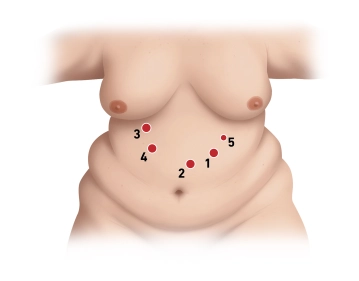
    Position the camera trocar in the left epigastrium, approximately 2-3 fingerwidbreadths caudal to the left costal arch in the midclavicular line.

    Access is by means ofthrough an optical trocar (12 mm Xcel trocar) under camera view. During insertion, ensure that the individual tissue layers can be viewed.During insertion, optical trocars ensure a clear view of the individual tissue layers. Place the additionalother trocars under camera view and after placementbringing the patient into the anti-Trendelenburg position.

    Then place three more 12 mm trocars in the following order. First around 1.5 hand’s width caudal to the xiphoid process in the left paramedian region (far supraumbilical), then approximately in the midclavicular line immediately at the right costal arch, introducing the tip of the trocar in the direction of the patient’s left leg.Introduce the first trocar about 1.5 hand widths inferior to the xiphoid process paramedian on the left (far supraumbilical) and the second trocar roughly in the midclavicular line immediately at the right costal arch, guiding the trocar tip in the direction of the patient's left leg. Finally,Now insert the third 12 mm trocar about one hand’s width caudalinferior to the right costal arch medial to the midclavicular line in the right upper quadrant.,then And finally insert an additional 5mm trocar at the left costal arch somewhat cranialsuperior to the first 12 mm trocar.

    Note: A liver retractor is usedneeded only if the liver isfor extremely large livers, which is the case in less than 5% of patients.

  • Dissection at the cardial notch

    Video
     

    Between the angle of His (without freely dissecting it completely) and somewhat medial to the upper pole of the spleen, detach the physiological adhesions between the gastric fundus and diaphragm or retroperitoneal space as much as possible using ultrasonic dissection.By ultrasonic dissection take down the physiological adhesions between the gastric fundus and diaphragm / retroperitoneal space as much as possible from the cardial notch (without freeing it completely) to somewhat medial to the superior pole of the spleen. This defines the cranialsuperior end of the stapled suture line.

    Note: Hiatal revision is not routinely done, but only in exceptional cases, e.g., in relatively low-weight patients with relatively low weight and substantial reflux problems. The surgeryprocedure results in creation ofcreates a low-pressure system by bypassing the pylorus and simultaneously applying tensiontraction on the pouch, thereby resulting asin a quasi-prophylactic measure against reflux. 

  • Opening the lesser sac at the superior aspect of the antrum

    Video
     

    After visualizingexposing the pylorus, enter the lesser bursasac at the superior edgeaspect of the gastric antrum approximatelyabout 2-–3  cm distal to the inferior borderaspect of the so-called “crow’s foot.” (branches of the vagi to the antropylorus). On the lesser curvature aspect, visualize the posterior wall of the stomach proximally and detach adhesions to the pancreas.Expose the posterior gastric wall proximally along the lesser curvature and take down adhesions with the pancreas.

  • Fashioning the gastric pouch I

    Video
     
    Paid content (text)
  • Fashioning the gastric pouch II

    Paid content (video)
    Paid content (text)
  • Opening the pouch

    Paid content (video)
    Paid content (text)
  • Measuring the biliopancreatic loop

    Paid content (video)
    Paid content (text)
  • Enterotomy

    Paid content (video)
    Paid content (text)
  • Transposition to the upper abdomen and gastroenterostomy

    Paid content (video)
    Paid content (image)
    Paid content (text)
  • Closing the anterior defect

    Paid content (video)
    Paid content (text)
  • Methylene blue test

    Paid content (video)
    Paid content (text)
  • Hemostasis at the staple line; drainage

    Paid content (video)
    Paid content (text)
date of publication: 18.03.2018

Single Access

Access to this lecture
for 3 days

€4.99 inclusive VAT

SEPA Maestro Mastercard VISA

webop-Account Single

full access to all lectures
price per month

for the modul: vascular surgery

from 8,17 €

hospitals & libraries

for the modul: vascular surgery

from 390,00 euros