Ileostomy, construction of - general and visceral surgery

You have not purchased a license - paywall is active: to the product selection
  • Stoma site marking

    Video
     
    Stoma site marking
     

    Ideally, the stoma should be marked and the subsequent instructions for stoma care given by specially trained stoma nurses or an experienced surgeon.

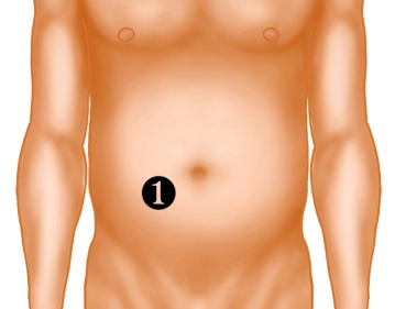
    1. Trial marking, with the patient supine or already sitting, within the right rectus abdominis (level of the umbilicus) in a 10×10cm skin area, preferably without folds and creases, scars and bony prominences.
    2. Check of the planned site with the patient in motion (standing, stooping down).
    3. The selected  site should be easily accessible to the patient and within his/her visual field and away from the natural beltline.
    4. To allow for intraoperative complications marking a secondary location is recommended.
    5.  Dressing the markings with sensitive skin bandages.

    The site of the ileostomy deeply affects its management and thus the patient’s quality of life!

  • Locating the loop of the terminal ileum

    Video
     
    Locating the loop of the terminal ileum
     

    After opening the abdominal cavity or completion of the major procedure, locate the loop of the terminal ileum with which to construct the loop ileostomy. The loop selected for the stoma should be at least 20cm proximal to the ileocecal valve.

  • Taping the ileum loop

    Video
     
    Taping the ileum loop
     

    Pass a silicone tape through a mesenteric window created close to the bowel preferably in an avascular area.

  • Skin incision for stoma and dissection down to the fascia.

    Video
     
    Skin incision for stoma and dissection down to the fascia.
     

    While lifting up the skin with, e.g., a towel clamp make a circular skin incision at the preoperatively marked site. The diameter of the skin incision should not exceed 3cm. Then dissect the subcutaneous tissue down to the fascia.

  • Dividing the anterior lamina of the rectus sheath

    Paid content (video)
    Paid content (image)

    After excision of skin and subcutaneous tissue, open up the anterior lamina of the rectus sheath in cruciform fashion. Stretching the fascia hereby will help avoid overlapping

  • Splitting the muscles

    Paid content (video)
    Paid content (image)

    Split the rectus muscle in the direction of its fibers until the posterior lamina of the rectus sheath is exposed.

  • Dividing the posterior lamina of the rectus sheath and the peritoneum

    Paid content (video)
    Paid content (image)

    Also divide the posterior lamina of the rectus sheath, together with the peritoneum, in cruciform fashion creating an opening just large enough to pass three fingers through ("three-finger test").

  • Delivering the ileum through the abdominal wall

    Paid content (video)
    Paid content (image)

    Pass the tape through the abdominal wall trephine and deliver the bowel loop, with the proximal limb located inferiorly. Some surgeon secure the loop with a rod.

  • Enterotomy and eversion of the proximal limb

    Paid content (video)
    Paid content (image)

    After closing the laparotomy and sterile draping (to avoid fecal contamination), perform a transverse enterotomy, e.g., with diathermy. Do this asymmetrically centered on the distal limb because this will evert the proximal limb which then is sutured to the skin.

  • Suturing the ileum to the skin

    Paid content (video)
    Paid content (image)

    Suture the bowel to the skin with interrupted monofilament sutures (e.g., PGA) 3/0. The inferior (proximal) limb is markedly everted and then secured to the skin in order to prevent contact of the aggressive intestinal juice with the skin.

  • Stoma care

    Paid content (video)
    Paid content (image)
    Paid content (text)