
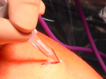
Skin incision left subcostal in the midclavicular line. Insertion of the Veress needle after local infiltration anesthesia; safety tests and establishment of the capnoperitoneum.
-
Skin Incision and Insertion of the Veress Needle
![Skin Incision and Insertion of the Veress Needle]()
Soundsettings -
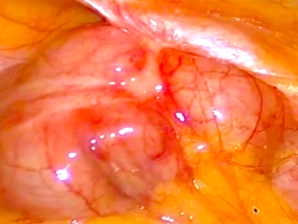
Insertion of the 10 mm trocar; diagnostic laparoscopy
![Insertion of the 10 mm trocar; diagnostic laparoscopy]()
Soundsettings Removal of the Veress needle; spreading of the muscle using scissors and blunt insertion of the 10 mm trocar.
Laparoscopic exploration: Mild fibrotic changes on the liver surface; the cecum carcinoma is adherent in a small area to the parietal peritoneum of the lateral abdominal wall. -
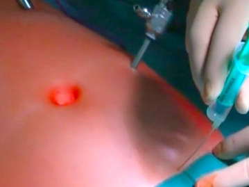
Insertion of the 5 mm Trocars; Positioning
![Insertion of the 5 mm Trocars; Positioning]()
Soundsettings Insertion of two 5 mm trocars in the left mid-abdomen in the midclavicular line and in the left lower abdomen paramedian – each under diaphanoscopy, local infiltration anesthesia, and skin incision – under direct vision. Trendelenburg positioning and left tilt of the table, shifting the small intestine to the left upper abdomen.
-
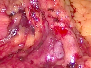
Preparation of the Ileocolic Vessels
![Preparation of the Ileocolic Vessels]()
Soundsettings Tensioning of the mesentery at the ileocecal junction and thus locating the ileocolic vessels. Dissection of the origin of the ileocolic artery from the superior mesenteric artery. Also, dissection of the venous origin.
-
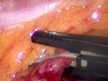
Transection of the Ileocolic Artery and Vein
![Transection of the Ileocolic Artery and Vein]()
Soundsettings Transect the ileocolic artery between Lapro-Clips® after additional sealing of the central vessel using LigaSure™ V. Subsequently, also transect the corresponding vein with sealing using LigaSure™ V.
-
Preparation of Mesocolon and Mesentery
![Preparation of Mesocolon and Mesentery]()
Soundsettings Layered dissection of the mesocolon or mesentery from the retroperitoneum with visualization and preservation of the duodenum, respecting the Gerota's fascia as the boundary layer. Determination of the oral resection margin.

Mobilization of the Terminal Ileum
Releasing the mesenteric adhesions of the terminal ileum from the lateral side for complete mobiliz
Releasing the mesenteric adhesions of the terminal ileum from the lateral side for complete mobiliz
Activate now and continue learning straight away.
Single Access
Activation of this course for 3 days.
US$9.30
inclusive VAT
Most popular offer
webop - Savings Flex
Combine our learning modules flexibly and save up to 50%.
from US$7.26 / module
US$87.13/ yearly payment
general and visceral surgery
Unlock all courses in this module.
US$14.52
/ month
US$174.30 / yearly payment
Webop is committed to education. That's why we offer all our content at a fair student rate.