Start your free 3-day trial — no credit card required, full access included

Anatomy - Right hemicolectomy, laparoscopically assisted

  1. Overview

    Overview

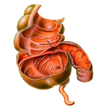
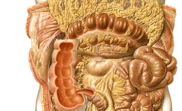
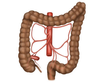
    The colon frames the inner abdominal wall and runs below the liver and stomach, surrounding the loops of the small intestine. The position of the colon is intra- or secondarily retroperitoneal. Its primary function is the thickening of the chyme through the absorption of water. The total length of the colon averages 120-150 cm. The colon begins at the ileocecal valve and ends at the rectosigmoid junction, where it transitions into the rectum.

    The colon is divided into the following sections:

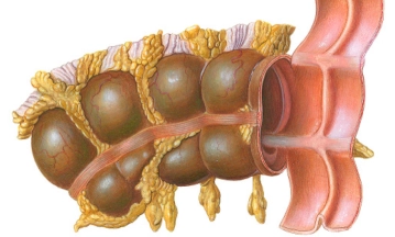
    • Cecum with the appendix
    • Ascending colon
    • Transverse colon
    • Descending colon
    • Sigmoid colon
  2. Macroscopy

    Macroscopy
    • The longitudinal muscle layers of the colon form three band-like muscle strips that are visible from the outside and are referred to as taeniae. They are distinguished as follows:
    • Taenia mesocolica: located towards the mesocolon
    • Taenia libera: free on the surface, facing the abdominal wall
    • Taenia omentalis: connected with the greater omentum
    • Appendices epiploicae are fat appendages from the tela subserosa in the area of the free taeniae.
    • Plicae semilunares are indentations of all wall layers intraluminally, whereas haustra are the outward bulges between them.

    For right hemicolectomy, the following intestinal sections are relevant from oral to aboral:

    • Right flexure and right lateral hemicolon
    • Ascending colon
    • Cecum with vermiform appendix
  3. Cecum

    Cecum
    • Definition: blind-ending initial part of the ascending colon, corresponding to an outpouching of the same; lat.: caecus = blind
    • Length 5-7 cm
    • Synonyms: Cecum, Blind gut
    • Location: intraperitoneal or also secondarily retroperitoneal, below the ileocecal valve, the Valva Bauhini
    • Identification: by the taeniae and the vermiform appendix
    • Vermiform appendix: intraperitoneal, in the area of the converging taeniae at the cecal pole, length: 6-20 cm, on average 8 cm, at the origin of the appendix the 3 taeniae converge
  4. Ascending Colon

    Ascending Colon
    • Definition: Section of the large intestine between the ileocecal valve, the entry of the small intestine (ileum), and the right colic flexure
    • Synonyms: ascending colon, or simply Ascendens
    • Location: [secondarily] retroperitoneal, extends from the right lower abdomen to the right flexure – peritoneal fixation to the posterior abdominal wall by the so-called Toldt's fascia
    • Length: 12-20 cm
    • Identification: by the longitudinal muscle fiber bundles – the taeniae, which retain their position – designations from the transverse colon; the transverse diameter is significantly larger than that of the descending colon!
Transverse Colon

Synonyms: transverse colon or simply Transversum.Location: intraperitoneal; mobile fixation through

Activate now and continue learning straight away.

Single Access

Activation of this course for 3 days.

US$9.40  inclusive VAT

Most popular offer

webop - Savings Flex

Combine our learning modules flexibly and save up to 50%.

from US$7.29 / module

US$87.56/ yearly payment

price overview

general and visceral surgery

Unlock all courses in this module.

US$14.59 / month

US$175.10 / yearly payment

to top