Right hemicolectomy

  1. Laparotomy

    Video
    Laparotomy
    Soundsettings
    The video clip can be played back with the automatic soundtrack of the subtitles.

    In the sidebar registered users can enable and disable the automatic start of the dubbing.

    Remark: Since this is a text-to-speech computer voice, it may mispronounce some medical terminology.

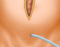
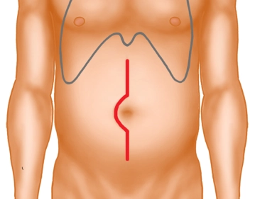
    Median midline laparotomy with right-sided umbilical incision. Transection of subcutaneous fat tissue and fascia with monopolar diathermy.

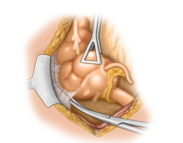
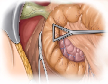
    After transection of the peritoneum, insertion of a suprapubic bladder catheter. Positioning of the surgical site with the retractor system.

    69-5
  2. Mobilization of the ascending colon

    Video
    Mobilization of the ascending colon
    Soundsettings
    The video clip can be played back with the automatic soundtrack of the subtitles.

    In the sidebar registered users can enable and disable the automatic start of the dubbing.

    Remark: Since this is a text-to-speech computer voice, it may mispronounce some medical terminology.

    The preparation of the right hemicolon is performed from lateral to medial and begins with the incision of the peritoneum lateral to the colon. This allows direct access to the slit-like space between the parietal fascia covering the retroperitoneal organs and the mesenteric/visceral fascia that extends continuously over all intraperitoneal organs.

  3. Preparation of the mesenteric root

    Video
    Soundsettings
    The video clip can be played back with the automatic soundtrack of the subtitles.

    In the sidebar registered users can enable and disable the automatic start of the dubbing.

    Remark: Since this is a text-to-speech computer voice, it may mispronounce some medical terminology.

    From the dorsal side, the mesocolon is now mobilized with the entire mesenteric root towards the ventral side.

    By preparing along the dorsal leaf of the mesocolic fascia and strictly avoiding injury to the ascending mesocolon, the mesenteric root is reached under complete mobilization of the ascending colon. This allows controlled access to the superior mesenteric vein with the veins draining there and the origin of the colonic arteries.

  4. Kocher maneuver

    Video
    Kocher maneuver
    Soundsettings
    The video clip can be played back with the automatic soundtrack of the subtitles.

    In the sidebar registered users can enable and disable the automatic start of the dubbing.

    Remark: Since this is a text-to-speech computer voice, it may mispronounce some medical terminology.

    The mobilization plane extends along the dorsal layer of the mesocolon to beneath the duodenum with the pancreatic head, resulting in a Kocher maneuver. The duodenum and pancreatic head are also covered by the visceral fascia, the mesoduodenum, and mesopancreas. This mesoduodenum is separated from the parietal fascial layer, which covers the vena cava and aorta.

    Note: At the end of the video, the surgeon demonstrates the detachment of the mesenteric layer, here the mesoduodenum and mesopancreas, from the parietal layer covering the vena cava and aorta. 

Dissection of the duodenum from the mesenteric root

Next, the duodenum is dissected from the ascending mesocolon dorsally by detaching the dorsal fasci

Activate now and continue learning straight away.

Single Access

Activation of this course for 3 days.

US$9.40  inclusive VAT

Most popular offer

webop - Savings Flex

Combine our learning modules flexibly and save up to 50%.

from US$7.27 / module

US$87.34/ yearly payment

price overview

general and visceral surgery

Unlock all courses in this module.

US$14.55 / month

US$174.70 / yearly payment