1. Overview

    Overview

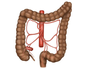
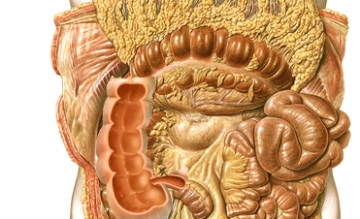
    The colon encircles the inner abdominal wall and runs below the liver and stomach, surrounding the loops of the small intestine. The position of the colon is intra- or secondarily retroperitoneal. Its primary function is the thickening of the chyme by absorbing water. The total length of the colon is on average 120-150 cm. The colon begins at the ileocecal valve and ends at the rectosigmoid junction, where it transitions into the rectum.

    The colon is divided into the following sections:

    • Cecum with the appendix
    • Ascending colon
    • Transverse colon
    • Descending colon
    • Sigmoid colon
  2. Macroscopy

    Macroscopy
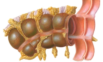
    • The longitudinal muscle layers of the colon form three band-like muscle strips that are visible from the outside and are referred to as taeniae. They are distinguished as follows:
    • Taenia mesocolica: located towards the mesentery
    • Taenia libera: free on the surface, facing the abdominal wall
    • Taenia omentalis: connected with the greater omentum
    • Fat appendages from the tela subserosa in the area of the free taeniae are referred to as appendices epiploicae.
    • Plicae semilunares are indentations of all wall layers intraluminally, whereas haustra are the outward bulges in between.

    For right hemicolectomy, the following intestinal segments are relevant from oral to aboral:

    • Right flexure and right lateral hemicolon
    • Ascending colon
    • Cecum with vermiform appendix
  3. Colon

    Colon
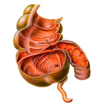
    • Definition: blind initial part of the ascending colon, corresponding to a bulge of the same; lat.: caecus = blind
    • Length 5-7 cm
    • Synonyms: cecum, blind gut
    • Location: intraperitoneal or also secondarily retroperitoneal, below the ileocecal valve, the Valva Bauhini
    • Identification: by the taeniae and the vermiform appendix
    • Vermiform appendix: intraperitoneal, in the area of the converging taeniae at the cecal pole, length: 6-20 cm, on average 8 cm, at the origin of the appendix the 3 taeniae unite
  4. Ascending colon

    Ascending colon
    • Definition: Large intestine section between the ileocecal valve, the entry of the small intestine (ileum), and the right colic flexure
    • Synonyms: ascending colon, or simply Ascendens
    • Location: [secondarily] retroperitoneal, extends from the right lower abdomen to the right flexure – peritoneal fixation to the posterior abdominal wall by the so-called Toldt's fascia
    • Length: 12-20 cm
    • Identification: by the longitudinal muscle fiber bundles – the taeniae, which retain their positional names from the transverse colon; the transverse diameter is significantly larger than that of the descending colon!
Transverse colon

Synonyms: transverse colon or simply Transversum.Location: intraperitoneal; mobile fixation by its

Activate now and continue learning straight away.

Single Access

Activation of this course for 3 days.

US$9.40  inclusive VAT

Most popular offer

webop - Savings Flex

Combine our learning modules flexibly and save up to 50%.

from US$7.27 / module

US$87.34/ yearly payment

price overview

general and visceral surgery

Unlock all courses in this module.

US$14.55 / month

US$174.70 / yearly payment