•Left colic flexure fixed to the diaphragm by the phrenicocolic ligament
•secondary retroperitoneal position of the descending colon
•Transition descending colon – sigmoid colon in the left iliac fossa
•intraperitoneal position of the sigmoid colon (→ sigmoid mesocolon)
•Transition sigmoid colon – rectum in front of the 2nd-3rd sacral vertebra
-
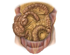
Descending colon and sigmoid colon
![Descending colon and sigmoid colon]()
-
Rectum
The rectum is divided into thirds. The height of the boundaries is measured with the rigid endoscope and the anocutaneous line is used as reference. Lower third 0-6cm, middle third 6-12cm, upper third 12-16cm.
-
Fascial Systems
•Parietal pelvic fascia covers the pelvic wall with vessels, autonomic nerves, and presacral venous/nerve plexus.
•Above the anorectal junction, meeting of the rectosacral fascia and the visceral pelvic fascia.
•The visceral fascias include the fascia propria of the pelvic organs (enclose the mesorectum dorsally and laterally) and the ventrally located Denonvilliers' fascia. -
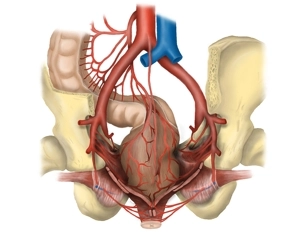
Vessels
![Vessels]()
•Anastomosis between the vascular territory of the superior mesenteric artery (A. colica media) and inferior mesenteric artery (A. colica sinistra) near the left colic flexure (arcade of Riolan).
•Supply of the left hemicolon, sigmoid colon, and upper rectum by the inferior mesenteric artery and its branches: A. colica sinistra, Aa. sigmoideae with the arcade of Drummond, A. rectalis superior.
•Supply of the upper third of the rectum via A. rectalis superior, which divides dorsally into two terminal branches, of the middle third from the A. rectalis media (paired, each as a branch of the A. iliaca interna) and of the lower third via A. rectalis inferior (paired, each as a branch of the A. pudenda interna from the A. iliaca interna). The Aa rectales mediae run in the so-called lateral ligaments of the rectum and are transected during total mesorectal excision.
•Venous drainage of the left hemicolon via veins of the same name into the portal vein territory.
•Venous drainage of the upper two thirds via the V. mesenterica inferior (portal vein territory) and of the lower third via the drainage area of the V. cava inferior.
Lymphatic drainage
•For all rectal segments along the course of the superior rectal artery and the inferior mesenteric
•For all rectal segments along the course of the superior rectal artery and the inferior mesenteric
Activate now and continue learning straight away.
Single Access
Activation of this course for 3 days.
US$9.30
inclusive VAT
Most popular offer
webop - Savings Flex
Combine our learning modules flexibly and save up to 50%.
from US$7.26 / module
US$87.13/ yearly payment
general and visceral surgery
Unlock all courses in this module.
US$14.52
/ month
US$174.30 / yearly payment
Webop is committed to education. That's why we offer all our content at a fair student rate.