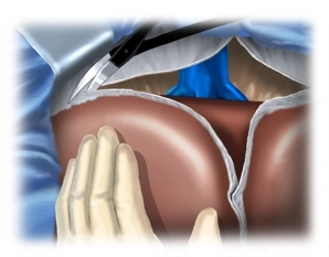
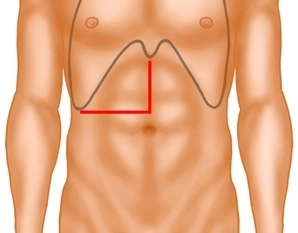
Left lateral liver resection, open - general and visceral surgery
You have not purchased a license - paywall is active: to the product selection
Laparotomy and exploration of the abdominal cavity
For the laparotomy the median incision is extended into the right flank just above the umbilicus Transect the right rectus muscle with bipolar scissors and incise the peritoneum. The incision may be kept to the left of the xyphoid: This yields good distance which facilitates the view, particularly regarding the hepatic veins.
Now transect the falciform ligament close to the abdominal wall. After lining the wound edges with towels insert the retractor for the abdominal wall and inspect the field: In primary cancer of the liver rule out extrahepatic metastasis and noticeably large lymph node in the hilum; in the example demonstrated here there is cirrhosis of the liver
Note:
Smaller procedures on the left hepatic globe, up to and including left lateral resection, may also be performed via median laparotomy, and wedge excisions of the inferior segments IVb, V and VI via a subcostal incision.
Freeing the liver
Start freeing the liver by transecting the left triangular ligament (bipolar scissors). Now free the liver from any adhesions with the diaphragm and expose the suprahepatic segment of the vena cava.
Note:
1. Careful exploration includes bimanual palpation of the liver which therefore must be fully freed.
2. Completely freeing the liver also helps to control possible bleeding complications.
3. Tumor invasion of the diaphragm does not contraindicate resection. The involved part of the diaphragm is resected en blocwith the tumor. In almost all cases the defect can be closed directly.
4. Since the left lobe of the liver is easily accessible, transection of the left triangular ligament is not mandatory.
Resecting the gallbladder
Started the resection by incising the serosa covering the anterior aspect of the hepatoduodenal ligament. Follow this by taking down the gallbladder in antegrade fashion from its hepatic bed to the hepatoduodenal ligament. Transect and ligate the cystic artery between Overholt dissecting forceps. This step in the dissection ends with exposure of the cystic duct.
Dissecting the hepatoduodenal ligament and lymph nodes
Hilar dissection
Local findings: Intraoperative ultrasonography (IOUS)
Dissecting the segmental hila, ligating the segmental artery II/III
Transecting the portal vein supply
Parenchymal dissection and bile duct transection
Dividing the left hepatic vein
Specimen retrieval and hemostasis
Checking for bile leakage, resecting the gallbladder
Covering the resection area with the falciform ligament
Closing the abdominal wall
Single Access
Access to this lecture
for 3 days
€4.99 inclusive VAT

webop-Account Single
full access to all lectures
price per month
for the modul: vascular surgery
from 8,17 €
Medical Equipment
Category | Medical Equipment | Company |
---|---|---|
Surgical instruments | BiClamp® 201 T ![]() | Erbe Elektromedizin GmbH ![]() |
Surgical instruments | Wasserstrahl-Applikator, gerade, mit monopolarer HF-Funktion ![]() | Erbe Elektromedizin GmbH ![]() |