Start your free 3-day trial — no credit card required, full access included

Anatomy - Anastomosis technique, biliodigestive, according to Hepp-Couinaud

  1. Liver and Biliary Tract

    Liver and Biliary Tract

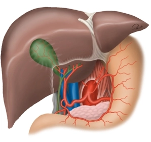
    The bile duct transports bile from the liver to the duodenum. In this way, bile contributes to the digestion of fatty foods. The bile ducts begin intrahepatically with the right and left hepatic ducts (ductus hepaticus dexter et sinister), which descend from the liver. When these two ducts meet, they form the common hepatic duct. As this duct continues to the duodenum, it is joined by the cystic duct, which comes from the gallbladder (Vesica biliaris). Together, they form the common bile duct, which opens into the duodenum. The major duodenal papilla (Vater's papilla) acts as a sphincter that regulates the flow of bile from the common bile duct into the duodenum.

  2. Jejunum

    • The jejunum is one of the three sections of the small intestine. It follows the duodenum and transitions into the ileum. Its German name "Leerdarm" is due to the fact that it is usually empty after death.*

    The jejunum is arranged in numerous loops and is attached to the dorsal abdominal wall by the mesentery. The corresponding section of the mesentery is called the mesojejunum; in conjunction with the ileum that follows the jejunum, the supporting structure is referred to as the radix mesenterii, which begins at the duodenojejunal flexure and ends at the ileocecal valve.

    • The jejunal loops are very mobile and thus variable in their position. While the beginning of the jejunum is clearly defined by the duodenojejunal flexure at the level of the second lumbar vertebra, the boundary between the jejunum and ileum cannot be sharply delineated anatomically. Like all hollow organs, the jejunum is lined with a mucous membrane (tunica mucosa). Externally, it is surrounded by a double layer of smooth muscle (tunica muscularis), and the outer organ is covered by the serosa of the peritoneum.
    • The upper part of the radix mesenterii carries vessels of the superior mesenteric artery to supply the jejunum, ileum, cecum, ascending colon, and the first two-thirds of the transverse colon.
    • The blood supply of the jejunum is provided by the jejunal arteries from the superior mesenteric artery. The veins of the jejunum drain into the superior mesenteric vein, which runs to the right of the superior mesenteric artery, then passes behind the head of the pancreas to join the portal vein.
    • The neural control of the jejunum is primarily mediated by the enteric nervous system. The regulation of peristalsis is carried out by the myenteric plexus (Auerbach's plexus), and the innervation of the mucosa by the submucosal plexus (Meissner's plexus). Additionally, the jejunum is regulated by the sympathetic and parasympathetic nervous systems (specifically the vagus nerve).

    from Latin ieiunus: fasting, hungry, lean *