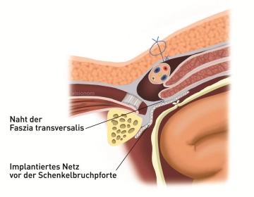
Femoral hernia repair – TIPP technique - general and visceral surgery

You have not purchased a license - paywall is active: to the product selection
  • Principle

    Principle
     

    The principle of this procedure involves closing the femoral hernia orifice with a synthetic mesh implanted via inguinal access. This requires division of the transversalis fascia which is closed subsequently with a simple Shouldice suture.

  • Skin incision

    Video
     
    Skin incision
     

    After local anesthesia perform a transverse skin incision of about 4 cm one finger width superior to the inguinal ligament. Transect the subcutaneous tissue and expose the aponeurosis of the external oblique.

  • Splitting the aponeurosis of the external oblique and snaring the round ligament of uterus

    Video
     
    Paid content (image)

    Now longitudinally divide the fibers of the external oblique, including the superficial inguinal ring. Clamp the edges of the fascia, pull up and dissect off the internal oblique. Expose and snare the round ligament of uterus.

  • Splitting the transversalis fascia and exposing the hernial orifice

    Paid content (video)
    Paid content (image)

    Split the transversalis fascia. Reduce the hernial sac from the gap between Cooper ligament and inferior margin of inguinal ligament. Bluntly dissect the peritoneum off the anterior pelvic wall.

    Ligate and resect any preperitoneal lipoma encountered here.

  • Trimming and implanting the mesh anterior to the hernial orifice

    Paid content (video)
    Paid content (image)

    Trim the mesh (in this case an adhesive model) to the appropriate size and implant it such that it will cover the posterior aspect of the hernial orifice. In the video clip note that the usual line of view, “from slightly anteroinferior“, is quickly changed to “from anterosuperior“, in order better view the implanted mesh deep down.

    Note:

    In the case demonstrated here the mesh is self-adhesive; otherwise fixate the mesh to the Cooper ligament with a few sutures.

  • Suturing the transversalis fascia

    Paid content (video)
    Paid content (image)

    Close the transversalis fascia with a running suture Prolene 2/0.  Another option (see figure) would be a double layered Shouldice suture.

  • Suturing the aponeurosis of the external oblique, subcutaneous tissue and skin

    Paid content (video)
    Paid content (image)