
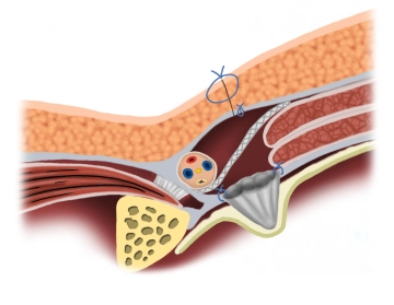
The Rutkow principle of inguinal hernia repair involves the insertion of an umbrella-like mesh plug into the local defect beneath the transversalis fascia. A second flat mesh, the onlay patch (similar to the Lichtenstein principle), is placed over the plug.
-
Principle
![Principle]()
-
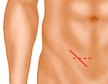
Inguinal skin incision
![Inguinal skin incision]()
Soundsettings A skin incision approximately 4 cm long is made obliquely 2 fingerbreadths above the inguinal ligament (solid line) or alternatively a transverse skin incision 2 fingerbreadths above the pubic bone. Subsequently, the subcutis is incised down to the external aponeurosis.
-
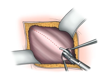
Splitting of the external aponeurosis
![Splitting of the external aponeurosis]()
Soundsettings The external aponeurosis is opened parallel to the fibers, including the external inguinal ring. The fascial edges are clamped, elevated, and the fascia is bluntly separated from the internal oblique muscle and the cremaster muscle.
Attention: The iliohypogastric nerve lies on the internal oblique muscle!
Exposure and looping of the spermatic cord
The spermatic cord is, if necessary, initially prepared and looped together with an indirect hernia
The spermatic cord is, if necessary, initially prepared and looped together with an indirect hernia
Activate now and continue learning straight away.
Single Access
Activation of this course for 3 days.
US$9.10
inclusive VAT
Most popular offer
webop - Savings Flex
Combine our learning modules flexibly and save up to 50%.
from US$7.08 / module
US$85.05/ yearly payment
general and visceral surgery
Unlock all courses in this module.
US$14.17
/ month
US$170.10 / yearly payment
Webop is committed to education. That's why we offer all our content at a fair student rate.