
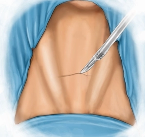
It is made caudal to the thyroid cartilage and approximately 1-2 fingerbreadths above the jugular fossa as a roughly 3 cm long transverse skin incision. Then, division of the subcutis and platysma to the superficial cervical fascia.
-
The Skin Incision
![The Skin Incision]()
Soundsettings -
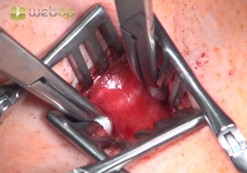
Access to the Trachea
![Access to the Trachea]()
Soundsettings After transverse transection of the superficial cervical fascia, a median incision of the pretracheal lamina and dissection into the depth. With two wound retractors, the ventral tracheal wall in the area of the third and fourth tracheal rings is optimally positioned, with the straight neck muscles being spread apart; if necessary, the thyroid isthmus must be pulled cranially.
Note: If the thyroid isthmus does not release the anterior tracheal wall, it is resected and the resection margin is sutured around both thyroid lobes.
The Opening of the Tracheal Window
The opening of the trachea is preferably performed between the second and third tracheal cartilage
The opening of the trachea is preferably performed between the second and third tracheal cartilage
Activate now and continue learning straight away.
Single Access
Activation of this course for 3 days.
US$9.20
inclusive VAT
Most popular offer
webop - Savings Flex
Combine our learning modules flexibly and save up to 50%.
from US$7.11 / module
US$85.43/ yearly payment
general and visceral surgery
Unlock all courses in this module.
US$14.23
/ month
US$170.90 / yearly payment
Webop is committed to education. That's why we offer all our content at a fair student rate.