Anatomy - Perineal rectosigmoidectomy – Altemeier procedure

  1. Pelvic floor

    Pelvic floor

    (1) Rectogenital septum, (2) Rectal stalks (paraproctium), (3) Mesorectal fascia

    Acting as a "functional unit" the muscular pelvic floor closes off the inferior abdominal cavity and prevents the prolapse of intestines and pelvic organs. The structures of the pelvic floor support the intestinal and urogenital outlet system, with the muscular parts playing an essential functional role in retention and continence. Muscular overstretching and denervation may result in pelvic floor descent or the various forms of lesser pelvis organ prolapse. Women are affected far more frequently than men (ratio 9: 1).

    Usually, the development of pelvic floor deficiency is a multifactorial process. The number of vaginal deliveries, excessive pressing during defecation, obesity, and previous pelvic surgery are some of the most common causes of pelvic floor disorders.

  2. Rectum

    Rectum

    From a functional point of view, the rectum is a storage reservoir for feces preventing its continuous discharge. The rectum directly adjoins the sigmoid colon and resembles it in that it has an S-shaped anteroposterior and lateral curvature (sacral, anorectal and sacral flexure). Typically, the rectum is divided into three segments:

    1. Rectal ampulla (main reservoir, anterior to the sacrum)

    2. Anal canal (see below, sealing zone between the anorectal flexure and the cutaneous orifice)

    3. Between 1. and 2. an unspecified section which emerges perpendicularly from 1. and joins 2., also at right angles. Here, the mucosa presents with typical folds (Kohlrausch's folds).

    Posteriorly the rectum is related to the sacrum and coccyx, laterally to the internal iliac arteries and veins, regional lymph nodes, sacral plexus and parts of the autonomic nervous system, as well as both ureters and uterine adnexa. In the female, the uterus and vagina lie anteriorly, while in men this is true for the bladder and prostate/seminal vesicles. Inferiorly the rectum is related to the pelvic floor.

  3. Anal canal

    Anal canal

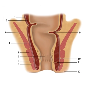
    (1) Tunica muscularis, longitudinal sublayer, (2) Tunica muscularis, circular sublayer, (3) levator ani, (4) puborectalis, (5) external anal sphincter, deep part, (6) anal columns, (7) external anal sphincter, superficial part, (8) external anal sphincter, subcutaneous part, (9) Kohlrausch’s fold, (10) internal anal sphincter, (11) proctodeal gland, (12) corrugator cutis ani

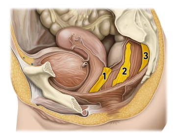
    The interaction of three muscles in the lower rectum creates a sphincter mechanism:

    1. The internal anal sphincter represents a thickening of the last annular fibers of the smooth colon muscles and is innervated by the sympathetic nervous system.

    2. The levator ani, however, has voluntary innervation (sacral plexus) and includes the puborectalis arising from the symphysis pubis. Since the course of the puborectalis creates a sling (deficient anteriorly) around the rectum, the latter becomes angulated.

    3. The external anal sphincter is also a striated muscle and extends from the center of the perineum (centrum perinei, perineal body) to the coccyx. Its somatic innervation is supplied by the pudendal nerve. With its contraction it completely seals off the anal canal.

    The different innervation of the three muscles involved in the sphincter mechanism provides additional protection against failure and resulting incontinence.

    The mucosa of the anal canal is plicated into numerous longitudinal folds (anal columns) displaying a dense arterial (!) plexus with venous drainage. When the sphincter muscles contract, these plexuses fill up quickly distending the mucosa and pushing the folds against each other, thereby ensuring a gas-tight seal. Hemorrhoids and venous thromboses are well known vascular complications in this region.

    Defecation involves not only relaxation of the sphincter mechanisms (initiated by voluntary muscle action, drainage of the cavernous bodies) but also active abdominal press and intestinal peristalsis.

Blood supply

Three main arteries supply the rectum with blood:Superior rectal artery (from the inferior mesenter

Activate now and continue learning straight away.

Single Access

Activation of this course for 3 days.

€4.99 inclusive VAT

Most popular offer

webop - Savings Flex

Combine our learning modules flexibly and save up to 50%.

from €3.29 / module

€39.50 / yearly payment

price overview

general and visceral surgery

Unlock all courses in this module.

€9.08 / month

€109.00 / yearly payment