Perioperative management - Rectovaginal fistula: Anterior levatorplasty and external sphincter plication

  1. Indications

    Indications

    Since spontaneous healing of rectovaginal fistulas is rather rare, even after fecal diversion by stoma, and conservative measures hardly ever lead to healing, the only adequate treatment is fistula surgery.

    Surprisingly enough, many patients present with long-standing fistulas, and this seems to be due to only minor or non-existent burden of suffering and, of course, shame. It is not unusual that the patients present for fistula repair at the urging of their partners or family; sometimes the diagnosis is made incidentally, e.g. in the context of diagnostic work-up and treatment of other disorders.

    At the core of the indication for surgical fistula repair are a significant degree of suffering, recurrent infections and any possible continence disorders. If the burden of suffering is not substantial enough and/or if the fistula is asymptomatic, the indication for surgery should only be governed by caution.

    When is the ideal time for fistula repair?

    Rectovaginal fistulas with a corresponding degree of suffering should be repaired as quickly as possible. However, the condition of the affected tissue plays a decisive role in the timing of the operation.

    Before initiating surgical treatment, any existing inflammatory processes, e.g. induration and inflammation, must have subsided as much as possible. If a fistula becomes symptomatic during abscess formation, the procedure is initially limited to abscess incision and flagging the fistula with a vessel loop, before undertaking the next step of repairing the fistula. Regular sitz baths, showering, possibly also debridement, a 10-14-day regimen of oral broad-spectrum antibiotics and a diet low in fiber may support the healing of inflammatory reactions.

    If the fistula was caused by perineal trauma, e.g. as a complication of the primary repair of a higher-grade perineal tear during vaginal delivery, the patient should wait at least 10-12 weeks after the initial event.

    When is a temporary diverting stoma indicated?

    Diverting ostomy is indicated in all types of fistulas with marked local inflammatory or scarring changes, extensive tissue defects, as well as anorectal incontinence requiring complex sphincter repair. So-called "complicated fistulas" involve:

    • Presence of several fistula openings
    • Horseshoe-shaped or suprasphincteric fistulas
    • Wide fistula openings, generally requiring difficult closure techniques (e.g. gracilis interposition)
    • Marked inflammatory perineal and perianal changes
    • Fistulas in Crohn’s disease
    • Radiation induced fistula
    • Severe obstetric injuries such as loss of perineum

    If a diverting stoma is required, the patient should receive detailed advice not only from the surgeon, but also from an ostomy nurse, as well as from self-help groups.

    Closure of a diverting stoma should take place no earlier than 3 months after successful fistula repair.

    Special case: Rectovaginal fistula in Crohn disease

    Fistulas developing in Crohn disease have an extremely poor prognosis with a recurrence rate of over 50%. Regardless of the location of the affected intestinal segment, fistula repair should not be undertaken in acute flare-ups. Surgical measures must be postponed until the episode has subsided, and the indication for a diverting fecal stoma should be broad.

  2. Contraindications

    Absolute contraindications

    • The general condition of the patient no longer permits a surgical procedure.
    • Acute flare-up in Crohn disease

    Relative contraindications

    • No burden of suffering
    • Asymptomatic fistulas


  3. Preoperative diagnostic work-up

    Preoperative diagnostic work-up

    The diagnosis of a rectovaginal fistula is primarily based on the patient's medical history and clinical examination.

    Medical history

    • Discharge of flatus or stool through the vagina (possibly only in diarrhea)
    • Fecal incontinence: Duration, type, frequency, influence of symptoms on everyday and social life
    • Relapsing inflammation of the vagina and lower urinary tract
    • Foul-smelling vaginal discharge
    • Vaginal deliveries
    • Maternal birth injuries: Perineal tear, episiotomy;
      (Caution: Sphincter lesions may also develop unnoticed during a seemingly "uncomplicated" delivery!)
    • Previous operations: Gynecology, urology, coloproctology
    • Psychosocial stress (subjective feelings, partner, family, job, leisure time)

    Clinical diagnostic work-up
    In order to rule out possible simultaneous causes of the existing incontinence, the diagnostic clinical work-up of rectovaginal fistulas must include the entire sphincter complex. Both the rectum and vagina must be inspected. The rectovaginal examination includes:

    • Inspection
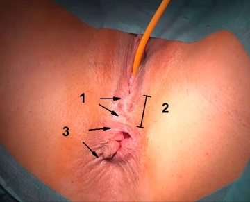
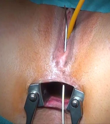
      Fistula openings, scars (see fig. 1), stool impurities or inflammatory vaginal changes, vaginal discharge; in fistulas after birth trauma with sphincter defect, the perineum is often narrow (see fig. 2), the anal mucosa is often tented toward the vaginal orifice, and the perianal skin has lost its characteristic rosette-like shape (see fig. 3)
    • Palpation
      A fistula opening at the vaginal orifice or in the rectum is best assessed and, if necessary, probed by bidigital examination.
    • Fistula probing
    • Proctosigmoidoscopy, colonoscopy

    Tricks to detect small caliber and high fistulas that are sometimes difficult to detect:

    • Rectoscopic insufflation of air → discharge via the vagina
    • Rectal application of blue dye diluted with normal saline with a bladder syringe (or H2O2, which prevents skin discoloration) → discharge via the vagina
    • Oral ingestion of poppy seed, which may be detected in the vagina with the help of a tampon (also useful in detecting vesicocolonic fistulas).

    Imaging
    Imaging modalities providing important additional information are used in unclear cases or complicated fistulas, e.g. in Crohn disease.

    Endoscopic ultrasound
    Transanal/endoanal ultrasound is the standard modality to be performed on each patient. The real-time mode allows assessment of both the internal and external anal sphincters and the levator sling. At the same time, it is possible to reveal other pelvic floor defects.

    Typically, endoscopic ultrasound will assess three regions:

    • Distal region → external anal sphincter
    • Middle region of anal canal → internal and external anal sphincter
    • Proximal segment → in addition the levator ani and pubococcygeus

    Magnetic resonance imaging (contrast-enhanced)

    • Detection of fistulas, abscesses and sphincter lesions

    Computed tomography

    • Less for fistula visualization than for ruling out concomitant pathologic processes (especially malignancies)

    Obsolete: Contrast-enhanced radiological fistula imaging

    Special case: Crohn disease

    In order to assess the activity of the underlying disease, which is critical to the time of surgery, Crohn patients should undergo staging prior to fistula repair:

    • Careful medical history
    • Abdominal ultrasound study
    • Colonoscopy
    • If necessary, examination under brief anesthesia to probe the fistula and insert vessel loops
    • If necessary, also gastroscopy and double contrast imaging of the small intestine
  4. Special preparation

    As a matter of principle, fecal contamination of the wound area during and after the procedure should be avoided:

    • Liquid diet 24 hours before surgery
    • Bowel cleansing as for colonoscopy or bowel resection, e.g. with oral preparations

    In addition:

    • Perioperative antibiotics: Cephalosporin + i.v. metronidazole
    • If necessary, preoperative construction of a diverting fecal stoma, see chapter on "Indications”
  5. Informed consent

    • Secondary bleeding
    • Local infections, abscesses
    • Continence disorders (increases with the number of previous operations/injuries)
    • Recurrent fistula
    • Wound dehiscence
    • Dyspareunia
    • Redo procedures
    • Positioning injuries (soft tissues, nerves)
    • Skin injury (due to electrocautery, disinfectants)
Anesthesia

General anesthesia Laryngeal mask airway anesthesia Spinal anesthesia ... - Operations in general,

Activate now and continue learning straight away.

Single Access

Activation of this course for 3 days.

€4.99 inclusive VAT

Most popular offer

webop - Savings Flex

Combine our learning modules flexibly and save up to 50%.

from €3.29 / module

€39.50 / yearly payment

price overview

general and visceral surgery

Unlock all courses in this module.

€9.08 / month

€109.00 / yearly payment