Gastrectomy - general and visceral surgery
You have not purchased a license - paywall is active: to the product selection
Bucket-handle incision
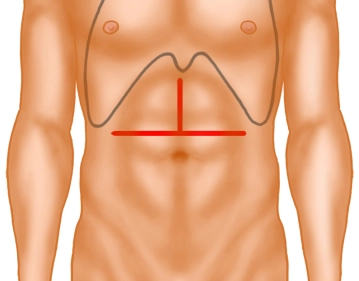
Open the abdomen via a transverse upper abdominal incision with upper midline extension, the so-called "inverted T".
Transect the subcutaneous tissue with electrocautery, tunnel both rectus muscles with a nonmetal probe and divide them with electrocautery.
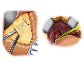
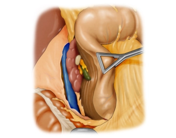
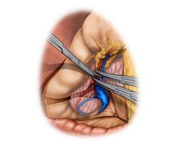
Explore the abdominal cavity to assess the location and extent of the tumor and thus its resectability after ruling out distant metastases, specifically in the peritoneum and liver. In this case the tumor is in the middle third of the stomach at the large curvature.
Dividing the lesser omentum, dissecting the gastroesophageal junction
After dividing the pericholecystic adhesions, transect the lesser omentum close to the liver in a distal to proximal direction while sparing the hepatoduodenal ligament. Continue the dissection to the gastroesophageal junction. Expose the full circumference of the abdominal esophagus and loop it with a tape.
Kocher maneuver
Follow this with the Kocher maneuver. To this end, incise the paraduodenal peritoneum about 1 cm from its lateral edge and in a largely avascular plane lift the duodenum from posterior away from the retroperitoneum and expose the vena cava.
Exposing the hepatoduodenal ligament
Dividing the greater omentum and gastroepiploic vessels
Freeing the greater omentum from the transverse colon will completely open the lesser sac. Leave the greater omentum attached to the large curvature. In the next step, divide the gastroepiploic vessels centrad between Overholt forceps.
Note: Complete mobilization of the stomach requires that later on the short gastric vessels from the gastric fundus to the spleen, as well as smaller vessels at the posterior wall of the stomach, a possible posterior gastric artery, and also the dense part of the lesser omentum, be divided or ligated.
Transecting the duodenum
Now expose the full circumference of the duodenum about 3 cm distal to the pylorus and transect it with a linear stapler (not shown in the video). Secure the staple line of the duodenal stump with serosizing interrupted sutures.
Note: Tension-free, secure closure of the duodenal stump is only possible if the duodenum has been mobilized enough by the previous Kocher maneuver.
Lymphadenectomy along the hepatic artery
Continue the lymphadenectomy to the left along the adventitia of the looped common hepatic artery. The first vessel encountered is the left gastric vein (formerly known as vena coronaria ventriculi), which is ligated and divided. The final step reaches the celiac trunk, where the left gastric artery is divided close its origin.
Caution! Rule out beforehand any aberrant left hepatic artery or large branches thereof arising from the left gastric artery.
Continue the para-aortic lymphadenectomy on the right to the right crus of the diaphragm.
Lymphadenectomy along the splenic artery
Complete the lymphadenectomy on the superior aspect of the pancreas along the splenic artery to the splenic hilum. Lift the entire lymph node package, which remains attached to the specimen, en bloc off the superior aspect of the pancreas and both arteries (common hepatic artery and splenic artery).
Note: In the video the specimen is still in situ at the completion of the surgical step.
Transecting the specimen
After truncal vagotomy transect the esophagus openly and remove the specimen. In the video the specimen has been opened and enlarged. The video then demonstrates the surgical site after the specimen had been removed, with the common hepatic artery and splenic artery still looped.
Note: Intraoperative frozen section of the esophageal resection margin is mandatory.
Constructing the Roux limb
In the next step construct a jejunal Roux limb. Under transillumination identify the mesenteric vessels about 20 cm–30 cm distal to the duodenojejunal flexure. Fashion an asymmetrical vascular pedicle proximad, since the blood supply should be primarily from distad. Transect the jejunum with a linear stapler (GIA) and serosize the distal stump. Now pass this efferent jejunal limb of adequate length into the upper abdomen through a window in the mesocolon.
End-to-side esophagojejunostomy
Jejunojejunostomy
Closing the abdomen
webop-Account Single
full access to all lectures
price per month
for the modul: vascular surgery