Perioperative management - Gastrectomy

  1. Indications

    • Primary gastric cancer
    • Remnant gastric cancer
    • Individual in resectable metastases, local peritoneal carcinomatosis (P1), recurrent malignancies (gastric stump cancer)
    • Unless there are massive risk factors (previous disease or general condition of the patient, see contraindications), surgery is generally indicated if curative treatment and local resectability are feasible.
    • Pre-/perioperative chemotherapy is recommended in locally advanced tumors.
    • Palliative gastrectomy may be indicated in rare cases of palliation (e.g., bleeding, perforation, stenosis).

    In order to achieve tumor-free resection margins (R0), a proximal safety distance in situ at the stomach of 5 cm (Lauren intestinal type) or 8 cm (Lauren diffuse type) must generally be maintained, except in mucosal carcinomas (T1a N0 M0). Lymph node clearance of compartments I and II is called D2-LAD and is the standard lymphadenectomy in gastric cancer. It can be regarded as the gold standard.

    The extent of the resection (total versus subtotal gastrectomy) is determined by tumor location/spread and the safety margin required by the histologic type.

    Special case of early gastric cancer
    Early gastric cancer is defined as a tumor restricted to the mucosa and submucosa of the gastric wall irrespective of lymph node status, spread and distant metastasis. By definition the gastric tunica muscularis is tumor-free.

    Early cancer differs with respect to the rate of potential lymph node metastasis. Tumors already infiltrating the submucosa are not candidates for endoscopic treatment, since there is a 4%–20% probability of lymph node metastasis. 

    About 5% of patients present with early gastric cancer of the mucosal type (pT1m), where a curative approach by endoscopic resection is possible as the probability of lymph node metastasis is extremely low. These patients have an excellent prognosis with a five-year survival rate of > 90%.

    Indication for endoscopic resection
    Superficial gastric cancer limited to the mucosa (about 5% of patients) and meeting the following criteria can be treated by endoscopic resection:

    • Lesions < 2  cm in size in elevated tumors
    • Lesions < 1 cm in size in flat tumors
    • Histologic grade: Well or moderately differentiated (G1/G2)
    • No macroscopic ulceration
    • Invasion limited to mucosa

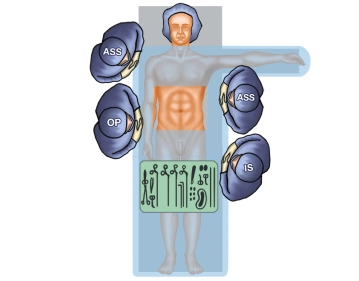
    The case presented here was diagnosed with a 5 cm large ulcerated gastric tumor at the large curvature in the transition zone from the middle to the distal third of the stomach. Histology demonstrated gastric cancer of the Lauren intestinal type, with endosonographic tumor staging yielding uT2, with no evidence of lymph node/distant metastasis. Therefore, indication for gastrectomy with D2 lymphadenectomy.

  2. Contraindications

    • Reduced general state of health (e.g., due to cancer cachexia)
    • Significant comorbidity with unfitness for anesthesia and/or surgery
    • Nonresectable tumor with proximal infiltration of the superior mesenteric artery or celiac trunk 
    • Any metastasis, except those cases where total R0 resection can be achieved (e.g., local peritoneal carcinomatosis, solitary liver metastasis, solitary distal lymph node metastasis).
    • Portal vein thrombosis and other disease with marked venous collateral circulation (liver cirrhosis)
    • In case of transit obstruction or tumor hemorrhage not controlled by endoscopy, gastrectomy may be indicated as a palliative procedure.
  3. Preoperative diagnostic work-up

    Mandatory:

    • Blood chemistry panel with so-called tumor markers (CA 72-4, CA 19-9, CEA)
    • Esophagogastroduodenoscopy with multilevel sampling (at least 8 biopsies)
    • Ultrasonography of abdominal cavity including the lesser pelvis; according to the German S3 guidelines the primary imaging modality when assessing liver metastases.
    • Endosonography to assess the T-stage, particularly to assess early gastric cancer (mucosa/submucosa type). Identification of risk constellations uT3/4 N+ and evidence of perigastric ascites. Lymph node assessment of category N1–2 is possible, but only with limited sensitivity and specificity. 
    • CT-study of chest and abdomen (with i.v. contrast agent)

    Optional:

    • Staging laparoscopy in advanced gastric cancer (cT3/cT4) to improve staging accuracy. It alone can detect previously unknown small subcapsular liver metastases and occult peritoneal carcinomatosis, possibly plus IOUS (intraoperative ultrasound) and  excision biopsy.
    • Peritoneal lavage with cytology is optional.
    • MRI should be reserved for those patients where computed tomography is not possible.
    • PET-CT is not routinely recommended in gastric cancer staging.
  4. Special preparation

    According to the German S3 guidelines, neoadjuvant chemotherapy is an integral part of the treatment strategy in gastric cancer.

    According to the current German guideline, diagnosed gastric cancer stage II and III (cT1/cT2N+ and cT3/resectable cT4Nx) should undergo perioperative chemotherapy. This is normally instituted before surgery (neoadjuvant) and continued postoperatively. This can increase the R0 resection rate, reduce the systemic recurrence rate and improve the overall prognosis.

    In planned preoperative chemotherapy (T3/4)

      1. Port implants
      2. Laparoscopic exploration optional
      3. Stenting in dysphagia, if needed

    In planned resection

    If necessary, initiate nutritional support before surgery in patients with marked preoperative weight loss.
    (on day before surgery)

      1. Nutrition: Clear broth
      2. Laxative measures 
      3. up to date blood chemistry panel, blood group and packed RBCs ordered
      4. Thrombosis prophylaxis (LMWH - low molecular weight heparin), compression stockings) (see guideline on thrombosis prophylaxis, section 1.10)
  5. Informed consent

    General surgical risks:

    • Thrombosis, pulmonary embolism
    • Pneumonia
    • Heparin induced thrombocytopenia (HIT)
    • Urinary tract infection

    Special surgical risks:

    • Mortality 2%–10%
    • Final decision regarding the actual resection procedure made intraoperatively
    • Injury to internal vessels and organs (incl. pleura!)
    • Suture line failure
    • Suture line failure of duodenal remnant
    • Endoluminal/ intraabdominal bleeding
    • Hypoperfused gastric reconstruction
    • Intraabdominal abscess, peritonitis
    • Pancreatitis and pancreatic fistulas
    • Secondary healing
    • Incisional hernia

    Possible need for extended surgery:

    • Cholecystectomy
    • Resection of liver metastases
    • Splenectomy

    Evidence of:

    • Temporary weight loss
    • Changing eating habits
    • Drains, gastric tube, urinary diversion
    • Possibly allogeneic blood transfusion
  6. Anesthesia

  7. Positioning

    Positioning
    • Supine
    • Right arm adducted
    • Left arm abducted
  8. Operating room setup

    Operating room setup
    • Surgeon on right side of patient
    • First assistant facing surgeon
    • Second assistant on right side craniad of the surgeon 
    • Scrub nurse on left side, caudad of first assistant
    • Possible third assistant on left side craniad of first assistant
  9. Special instruments and fixation systems

    • Gray Surgical Retractor System
    • Long instruments (including Allis clamps, Nakayama clamps, angled scissors)
    • Staplers (e.g., GIA 75, TA 90, EEA)
    • Coagulation, e.g., with argon beam coagulator, bipolar
  10. Postoperative management

    Postoperative analgesia: with epidurals, supportive i.v. analgesia

    Follow this link to PROSPECT (Procedures Specific Postoperative Pain Management)

    This link will take you to the International Guideline Library.

    Postoperative care: ICU monitoring (at least 24h); intra-abdominal target drains removed depending on flow rate; upper GI series on postoperative day 5 depending on patient condition; nasogastric tube removed if no leakage; stitches removed postoperative day 10–12; iron and vitamin replacement (vitamin B12, folic acid, vitamin D) during the later postoperative course (from about the third postoperative month).

    Deep venous thrombosis prophylaxis: Early ambulation, compression stockings, LMWH

    There is a high risk of thromboembolism due to the extensive surgery. Note: Renal function, HIT II (history, platelet check).

    This link will take you to the International Guideline Library.

    Ambulation: Physical rest until wound healing is complete.

    Physical therapy: Early ambulation, deep breathing exercises

    Diet: Parenteral nutrition and fluid replacement, enteral nutrition on postoperative day 1–4 via nasogastric tube, solid food only after upper GI series.

    Bowel movement:

    If bowel movement does not commence on its own after 3–4 days, a light laxative might be indicated.

    Work disability:
    Work disability usually lasts at least 4 weeks.