Start your free 3-day trial — no credit card required, full access included

Gastric bypass, laparoscopic

  1. Start of the Operation and Gastric Pouch Formation

    Video
    Start of the Operation and Gastric Pouch Formation
    Soundsettings

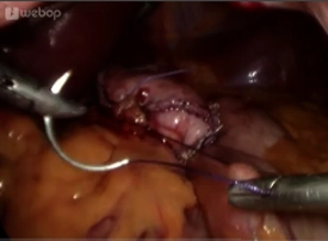
    After the start of the operation (establishment of the capnoperitoneum, placement of the working trocars, inspection of the abdominal cavity), the exposure of the angle of His follows.

    Subsequently, the formation of the gastric pouch begins in the area of the lesser curvature 2 cm below the esophagogastric junction. The left gastric artery supplies the gastric pouch and is taken into account to avoid bleeding complications and to ensure the blood supply of the pouch. The transection of the stomach is initially performed in a horizontal direction with linear stapling devices (staple height 3.5 mm).
    The dissection is then continued in a triangular shape towards the left diaphragmatic crus. The left crus of the diaphragm serves as the anatomical target line.
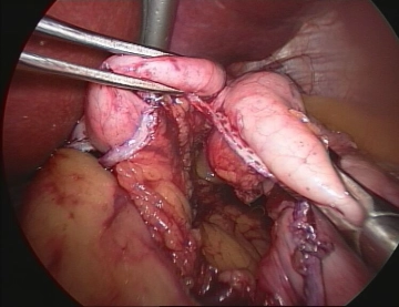
    The fundus should be completely severed to exclude the production site of the entero hormone ghrelin from the food passage and to avoid later dilatation.
    Finally, sufficient hemostasis at the staple line should be performed with ultrasound.

  2. Measurement of the biliopancreatic limb and elevation to the upper abdomen

    Soundsettings

    After complete transection of the stomach, the ligament of Treitz is sought. In case of a fat-rich greater omentum, it is necessary to transect it. Approximately 50 cm aboral from the ligament of Treitz, the jejunal limb is led antecolically up to the gastric pouch. It is helpful in measuring to consistently place the oral limb on the right in the image and fix it with the instrument of the right hand to avoid limb confusion. For measurement, the use of auxiliary instruments is useful.

    Alternative paths for the alimentary limb to the gastric pouch.

    Tip: To avoid the error of a too short limb, an initial check of the reachability of the pouch for the alimentary limb should be performed. If the limb is still too short, a primary sleeve gastrectomy-like pouch form and/or skeletonization of the alimentary limb or a retrocolic retrogastric access is recommended.

  3. Parallel Fixation of the Loop at the Gastric Pouch

    Parallel Fixation of the Loop at the Gastric Pouch
    Soundsettings

    The jejunal loop is attached to the gastric pouch tension-free with two fixation sutures.

    Note: If the loop is fixed too far cranially, it leads to the formation of a distal pouch reservoir with overflow phenomenon; if the fixation is too far caudally, the risk of dumping syndrome increases. Therefore, it should be applied at the second staple line row and adapted to the pouch shape and size.

    Tip: To avoid loop rotation (“blue loop” syndrome), the loop is brought up under vision (mesentery points to the left). If rotation occurs nonetheless, it is resolved and corrected.
    Also, loop confusion must be absolutely excluded, which requires consistent identification of the ligament of Treitz. A confusion must be corrected as an emergency, otherwise cardiac arrest threatens due to the overdistended stomach (distension).

  4. Creation of the antecolic-retrogastric gastroenterostomy (linear anastomosis of the posterior wall)

    Soundsettings

    For the creation of the gastroenterostomy, the gastric pouch and the alimentary loop are opened in parallel with the help of the ultrasound dissector. The gastric pouch is opened at the posterior wall.
    This can be done with an electric hook/scissors, ultrasound or Ligasure.
    The posterior wall of the anastomosis is closed using a linear stapler (blue magazine), after which the open anterior wall remains.

    Tip: To avoid a posterior perforation, the incision should be made carefully while simultaneously holding the fixation suture under tension and, in an emergency, oversewn.
    If a transection of the intestinal continuity has already occurred before the creation of the gastroenteroanastomosis, and the distance from the end of the alimentary loop is too large, a re-resection of the small intestine tip is performed.

  5. Creation of the Gastroenterostomy (Suture Closure of the Anterior Wall)

    Creation of the Gastroenterostomy (Suture Closure of the Anterior Wall)
    Soundsettings

    Before closing the stomach and intestinal openings, a calibration probe is inserted into the efferent jejunal limb.
    Subsequently, closure with a continuous suture and securing the anastomosis with additional single button sutures.
    Alternatively, the gastroenterostomy can also be created using a circular stapler or by complete hand suture.

    Tip: To avoid a postoperative anastomotic stenosis , an 8mm probe is placed over the anastomosis to prevent grasping the posterior wall during closure of the anterior wall.

  6. Testing of the Anastomosis

    Testing of the Anastomosis
    Soundsettings

    After completion of the anastomosis, the tightness of the anastomosis is checked by methylene blue test or by gas insufflation. Careful inspection from all sides must be observed. Subtle testing and rinsing with water is necessary to detect even small amounts of blue solution on the posterior wall.

    Note: The gastrojejunostomy is performed in primary procedures as a combined anastomosis with linear anastomosis of the posterior wall and suture closure of the anterior wall. In revision and switch operations, a complete hand-sewn anastomosis is performed.

Transection of the small bowel continuity/Division of the biliopancreatic limb

After opening a mesenteric window, division of the proximal small bowel limb with a stapler (blue c

Activate now and continue learning straight away.

Single Access

Activation of this course for 3 days.

US$9.30  inclusive VAT

Most popular offer

webop - Savings Flex

Combine our learning modules flexibly and save up to 50%.

from US$7.19 / module

US$86.33/ yearly payment

price overview

general and visceral surgery

Unlock all courses in this module.

US$14.38 / month

US$172.70 / yearly payment