Start your free 3-day trial — no credit card required, full access included

Appendectomy, laparoscopic

Reading time readingtime 16:32 min.
  1. Trocar positions

    54-01a-neu
    54-01b-neu

    The author presents two possible trocar arrangements. In the case demonstrated here, the 12mm trocar is placed at the navel, the 5mm in the midline suprapubic area, and a 10mm pararectal in the left lower abdomen.

    Alternatively, the 10 and 5mm trocars can be placed at the pubic hairline on the right and left.

  2. Instrumentation, Minilaparotomy

    Video
    Instrumentation, Minilaparotomy
    Soundsettings

    Infraumbilical incision, minilaparotomy, and insertion of the 12mm trocar under direct vision after placing fascial retention sutures.

  3. Pneumoperitoneum, Exploration

    Video
    Pneumoperitoneum, Exploration
    Soundsettings

    Creation of the pneumoperitoneum. The pressure plateau is selected according to the size, age, and gender of the patient (6-8 mmHg for children, 10-14 mm Hg for adults). The flow limitation of the CO2 gas should be set at 1 ml/min.

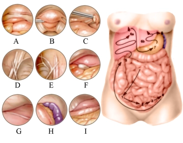
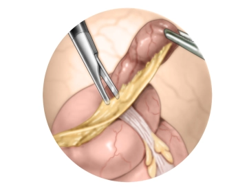
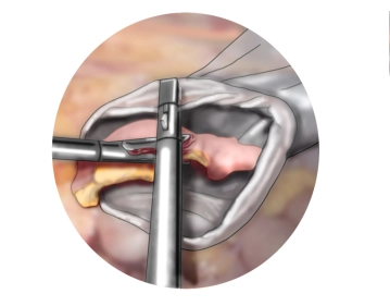
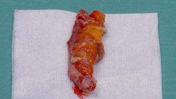
    Every laparoscopic procedure begins with the exploration of the abdominal cavity! Here, an acute purulent appendicitis is observed, which is inflamed and adherent to the anterior abdominal wall.

    In the film, the right lower abdomen is first explored, followed by the right upper abdomen, then the left upper abdomen, the right lower quadrant again, and finally the small pelvis.

    In relation to the sketch, the inspection of the lower abdominal organs occurs in numerical order, here after the inspection of the appendix area, uterus and bladder (A), the Douglas space (B), left ovary (C), and deep inguinal ring (D). It continues to the right over the right groin area (E). The optics are then directed to the right upper abdomen (5), inspection of the gallbladder (F) and right liver lobe (G). Further inspection of the left upper abdomen follows: spleen and stomach body (H) as well as the left liver lobe with falciform ligament, stomach, and greater omentum (I).

  4. Working trocars, mobilization of the appendix

    Video
    Working trocars, mobilization of the appendix
    Soundsettings

    Placement of a 10mm trocar pararectally and a 5mm trocar suprapubically under direct vision. Before each incision, local anesthesia is administered at the trocar insertion sites.

    The appendix is then bluntly mobilized with a swab, with tension applied to the appendix using a soft bowel clamp.


  5. Transection of the mesentery

    Video
    Transection of the mesentery
    Soundsettings

    Transection of the mesoappendix with monopolar until the base, clipping of the appendicular artery using titanium clips.

  6. Transection of the appendix

    Video
    Soundsettings

    Insert the Endo GIA (stapling device, 30 mm, blue cartridge) via a 12mm trocar and resect the appendix. Alternatively, appropriate appendix clips with locking can be used.

    Remark 1: Which method for laparoscopic resection of the appendix (Röder loop, clips, stapler…) is preferred cannot be answered even with the current meta-analyses, so cost-effectiveness will also play a role in decision-making.

    Remark 2: "Meckel" (searching for a Meckel's diverticulum) is only necessary with a relatively bland appendix. If a Meckel's diverticulum is found, it can be resected sufficiently far from its base with a stapling device (e.g., Endo GIA, 30 or 45 mm blue cartridge) (not shown in the film), as clear appendicitis is the cause of the symptoms.

  7. Retrieval of the appendix, swabbing of the Douglas pouch

    Video
    Retrieval of the appendix, swabbing of the Douglas pouch
    Soundsettings

    Transferring the appendix into a retrieval bag, then cleaning the Douglas pouch with one or more compresses introduced via a 12 trocar and checking for blood dryness. Depending on contamination, the paracolic gutter can also be swabbed.

  8. Extraction of the retrieval bag

    Video
    Extraction of the retrieval bag
    Soundsettings

    The retrieval bag is now removed through the channel of the 12mm trocar at the navel, requiring an extension of the fascia.

  9. Fascial closure, removal of trocars, skin suture

    Video
    Soundsettings

    After removal of the 12mm trocar, the fascia is closed infraumbilically (Vicryl 0, UR6 needle). This fascial closure is performed under laparoscopic control. Then, under camera view, the 5mm trocar is removed. Finally, the skin is closed with monofilament absorbable suture material (3-0/4-0).

    Note: In cases of a sanitized focus and local peritonitis, routine placement of a drain is not performed!

to top