Start your free 3-day trial — no credit card required, full access included

Anatomy - Ventral mesh rectopexy according to D'Hoore, laparoscopic

  1. Rectum

    Rectum

    Functionally, the rectum serves as a fecal reservoir, preventing continuous defecation. It directly follows the sigmoid colon and, like the latter, exhibits an S-shaped anterior-posterior and lateral curvature (sacral, anorectal, and lateral flexures). Typically, the rectum is divided into three sections:

    1. Ampulla recti (main reservoir, ventral to the sacrum)
    2. Anal canal (see below, closure region between the anorectal flexure and the skin opening)
    3. Between 1 and 2, an unspecified section that emerges at a right angle from 1 and transitions at a right angle into 2. Here, typical folds (Kohlrausch folds) are found in the mucosa.

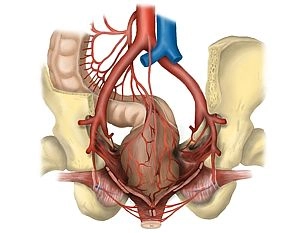
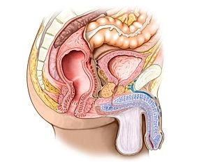
    Adjacent structures of the rectum include dorsally the sacrum and coccyx, laterally the regional lymph nodes next to the internal iliac artery and vein, the sacral plexus, and parts of the autonomic nervous system, as well as the ureter and adnexa. Ventrally, in females, are the uterus and vagina, and in males, the bladder and prostate/seminal vesicle. Caudally is the pelvic floor.

  2. Anal canal

    Three muscles form the anal sphincter complex in the wall of the lower rectum:

    1. The M. sphincter ani internus represents a thickening of the last circularly arranged fibers of the smooth colon musculature and is innervated by the sympathetic nervous system.
    2. The M. levator ani, on the other hand, is voluntarily innervated (sacral plexus) and includes the M. puborectalis attached to the pubic bone. It acts as a large loop around the anal canal anteriorly, functionally bending it.
    3. The M. sphincter ani externus is also striated and suspended between the center of the perineal region (Centrum perinei) and the coccyx. It is voluntarily innervated by the N. pudendus. Its contraction results in the terminal closure of the anal canal.

    The different innervation of the three closing muscles provides additional security against failures and resulting incontinence.
    In the mucosa of the anal canal, there are numerous longitudinal folds (Columnae anales) that contain dense arterial (!) plexuses with venous drainage. Upon contraction of the sphincter muscles, they quickly fill up, causing the mucosa to swell, adhere together, and thus create a gas-tight seal. Hemorrhoids and venous thromboses are known vascular-related complications of this region.

    Defecation occurs partly through the relaxation of the closure mechanisms (initiated by the voluntary muscles, emptying of the erectile tissue) and partly through active abdominal pressure and intestinal peristalsis.

Vascular supply

The rectum is supplied arterially by three main vessels:the superior rectal artery (from the inferi

Activate now and continue learning straight away.

Single Access

Activation of this course for 3 days.

US$9.40  inclusive VAT

Most popular offer

webop - Savings Flex

Combine our learning modules flexibly and save up to 50%.

from US$7.29 / module

US$87.56/ yearly payment

price overview

general and visceral surgery

Unlock all courses in this module.

US$14.59 / month

US$175.10 / yearly payment

to top