Start your free 3-day trial — no credit card required, full access included

Perioperative management - Right adrenalectomy, open

  1. Indications

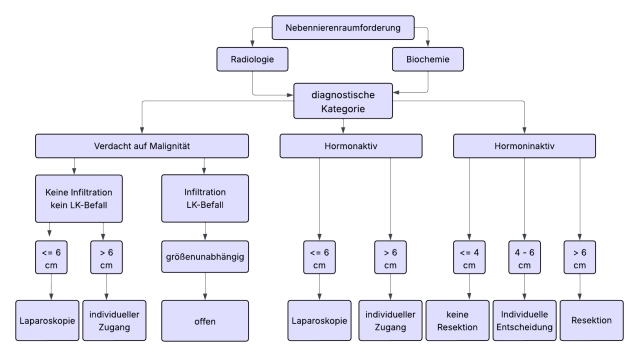
    There is consensus in the guidelines that most adrenal tumors should be operated on minimally invasively when indicated. Tumor diameter >6 cm and clear indications of malignancy in preoperative imaging are considered limits of minimally invasive surgery.

    For open adrenalectomy, the transabdominal or thoraco-abdominal approach is preferred, as the main indication for the open procedure is large tumors suspected of malignancy.

    For suspicious adrenal tumors of 6-8 cm in size and Hounsfield units (HU) (density values of the adrenal mass) >20, an open approach (+lymphadenectomy) is preferred without preoperative evidence of malignancy.

    An R0 en-bloc resection with surrounding retroperitoneal fat tissue should be aimed for, tumor capsule opening should be avoided, and at least morphologically conspicuous lymph nodes should be simultaneously removed.

    In the following situations, an indication for open adrenalectomy may exist:

    • Endocrine-active adrenal cortex tumors (Conn or Cushing adenomas, tumors with androgen secretion) especially with > 10 cm diameter

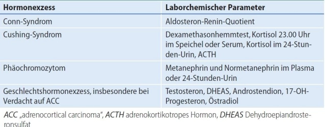
      • Conn syndrome (primary hyperaldosteronism, PHA):

        The most common cause of secondary hypertension is primary hyperaldosteronism.

        In the case of a unilateral aldosterone-producing adenoma, unilateral adrenalectomy is indicated.

        In patients with primary hyperaldosteronism (PHA) and bilateral adrenal changes, unilateral adrenalectomy may be considered if adrenal venous sampling (AVS; selective blood sampling from the adrenal veins) demonstrates clear functional lateralization. The significance of CXCR4-PET as a non-invasive alternative to AVS must be validated by further studies.
         
      • Cushing syndrome(hypercortisolism)

        A florid adrenal Cushing syndrome with classic clinical stigmata represents an indication for surgery.

        Before a possible surgical removal of the adrenal tumor, ACTH independence of cortisol excess must be confirmed to prevent the procedure from being mistakenly performed, although the cause of hormone excess is, for example, pituitary due to an adenoma of the anterior pituitary (central Cushing syndrome) or paraneoplastic syndrome with ectopic ACTH secretion in tumor disease.
         
      •  Sex hormone-producing adrenal cortex tumors

        An adrenocortical carcinoma is the most common cause of clinically relevant pathological androgen/estrogen secretion from the adrenal gland, adenomas are very rare.
         
    • Adrenocortical carcinoma Adrenocortical carcinoma (ACC) ENSAT stage I-III Link to ENSAT classification
      • At initial diagnosis, almost always > 4cm and exhibits endocrine activity in 50-80%. Typical is cortisol production or mixed hormonal production (androgens/estrogens and cortisol).
      • In adrenocortical carcinoma, open adrenalectomy is the gold standard. For tumors < 6 cm without evidence of local or lymph node infiltration (ENSAT Std. I+II), minimally invasive adrenalectomy can be performed.
      • A radical tumor resection with removal of the adrenal gland and all fat/connective tissue in the affected compartment without capsule opening and a lymphadenectomy, this with uncertain data situation, is recommended. A definition regarding the extent of the required lymphadenectomy is not yet available.
      • Note: The rate of local and peritoneal recurrences is increased in the laparoscopic group according to current evidence. Conversion from laparoscopic to open adrenalectomy worsens overall survival.
         
    • Pheochromocytoma (PC) Adrenal medulla tumor with catecholamine excess in cases of suspected malignancy, very large tumors, or when technical difficulties are expected.

      • Approximately 1/3 of all pheochromocytoma patients are associated with a hereditary tumor syndrome. Genetic screening is an indispensable part of pheochromocytoma diagnostics. SDHB-associated tumors have a particularly high risk of malignancy and recurrence.
      • Overall, 10% of PCs are malignant. Metastases are the only proof of a malignant pheochromocytoma.
         
    • Myelolipomas do not per se constitute an indication for surgery.

      Large adrenal myelolipomas (hormonally inactive benign tumors containing mature fat tissue and hematopoietic tissue) can become symptomatic through necrosis or spontaneous bleeding. In these cases, resection may be required.
       
    • Adrenal metastases (from malignant tumors of other origins) should be removed if no further metastases are present and tumor freedom can be achieved through removal.

      Note: Metastasis adrenalectomy should be performed minimally invasively, provided the metastasis can be removed in toto and without tumor cell dissemination. An open approach is reserved for the few cases where there is evidence of local infiltration or if the metastasis exceeds 6 cm.
       
    • Large schwannomas or adult neuroblastomas
       
    78-Nebennierenraumforderung
  2. Contraindications

    • Pulmonary or cardiac problems that exclude surgical intervention.
       
    • No adrenalectomy without prior determination of hormonal activity!
       
    • Due to the very low risk of malignancy, surgery is generally not indicated for hormonally inactive, non-malignant-suspect tumors < 4 cm. If surgery is performed, it must be justified.
       
    • In bilateral primary hyperaldosteronism (idiopathic adrenal hyperplasia) without lateralization, total adrenal removal is not indicated. Initiation of therapy with aldosterone antagonists.
       
    • No adrenalectomy but parenchyma-sparing adrenal resection (to preserve function, one-third of an adrenal gland should be left intact!).
      • In hereditary (bilateral) pheochromocytomas, as the likelihood of metachronous contralateral PC is high and the risk of malignancy is considered low.
      • In Cushing's syndrome based on bilateral macronodular hyperplasia. Balancing act between cortisol control and residual hormonal function. After total adrenalectomy, there is a requirement for substitution with the risk of a life-threatening Addisonian crisis (acute adrenal insufficiency).
         
    • Myelolipomas and adrenal cysts should only be removed if there are clinical symptoms due to compression of adjacent structures, necrosis, or spontaneous bleeding.
       
    • Other contraindications depend on underlying diseases (surgical risk) and the significance of adrenalectomy for the patient's quality of life or life expectancy.
  3. Preoperative Diagnostics

    Adrenal tumors include benign and malignant changes of the adrenal glands that affect morphology and/or function. The aim of diagnostics is to classify morphological and/or functional changes to evaluate therapeutic options.

    Diagnostics include taking a medical history and performing a physical examination, with blood pressure and heart rate measurement being mandatory components. Additionally, endocrinological methods and imaging techniques are required.

    Tumorous changes in the adrenal glands should always be evaluated for possible hormonal activity: catecholamines, aldosterone, cortisol, sex hormones, or their precursors.

    Clinical Presentation

    Typical leading symptoms in pheochromocytoma (PC) are severe hypertension, paroxysmal headaches, and palpitations. Pathognomonic for hypercortisolism (Cushing's syndrome) are central obesity, buffalo hump, moon face, striae rubrae, hirsutism, and plethora.

    Screening for primary hyperaldosteronism (PHA) in cases of hypertension or hypokalemia

    Laboratory Diagnostics/Hormonal Diagnostics:

    The initial screening tool for suspected autonomous cortisol secretion (ACS) is the dexamethasone suppression test. Based on the level of dexamethasone-suppressed serum cortisol, a distinction is made between a non-functional adrenal tumor and a "(possible) autonomous cortisol secretion" in clinically asymptomatic patients. A serum cortisol level above 5 ng/dl confirms the diagnosis. A low to suppressed ACTH (< 10 pg/ml) is necessary for the diagnosis to rule out pituitary hypercortisolism or ectopic ACTH production.

    Diagnostics for PC include the determination of metanephrines in plasma or 24-hour urine collection. A clonidine test may be considered if findings remain borderline.

    3-Methoxytyramine in plasma serves as a tumor marker for an SDHB mutation and is used as preoperative screening for metastases in a PC > 5cm.

    In primary hyperaldosteronism (PHA) (Conn's syndrome), there is autonomous, i.e., renin-independent aldosterone secretion. The screening parameter is the aldosterone-renin ratio (ARR), which can also identify patients with mild primary hyperaldosteronism. Aldosterone and renin are usually determined in the morning after 5 to 15 minutes of sitting. If the ratio is elevated, a confirmatory test is required according to guidelines, such as a saline infusion test (2l NaCl 0.9% i.v. over 4 hours). In healthy individuals, aldosterone suppression occurs after 4 hours, while it does not in affected individuals. The influence of antihypertensive medications on the RAAS (Renin-Angiotensin-Aldosterone System) must be considered.

    Sex hormones and steroid precursors in the clinical presentation of sex hormone excess (hirsutism, gynecomastia in men), tumor > 4 cm, or imaging suspicion of ACC: Steroid precursors (DHEA/DHEA-S, androstenedione, 17-α-hydroxyprogesterone), testosterone in women, 17β-estradiol in men and postmenopausal women.

    Note: Approximately 60 – 80% of adrenocortical carcinomas secrete hormones (functionally active), with the most commonly produced hormones being glucocorticoids (especially cortisol) and androgens.

    78-Labor hormonaktive NN Tumoren

    Genetic Testing

    Current guidelines recommend genetic testing for all patients with pheochromocytoma regardless of age, tumor number, or location.

    Note: Pheochromocytoma (PC) is an indicator lesion for hereditary syndromes. In unilateral cases, 12% have a hereditary condition, while in bilateral PC, young patients, or those with a notable family history, 30% have a hereditary disease.

    Imaging Techniques:

    Computed Tomography

    CT with and without contrast is recommended as the basic procedure for visualizing adrenal tumors. It allows for the determination of the size of the adrenal tumor and its fat content (Hounsfield units, HU), as well as an assessment of possible surrounding infiltrations.

    Native CT (Native CT with determination of the density values of the adrenal mass in Hounsfield units (HU))

    The native CT is the first important step in evaluating an adrenal tumor, especially for determining tissue density (HU).

    Lesions with < 10 HU can be classified as adenomas with a sensitivity of 70% and specificity of 95% and are primarily not suspected to be malignant.

    Contrast-enhanced CT is used additionally when the diagnosis in the native CT is not clear.

    Dynamic contrast-enhanced CT with determination of the so-called "washout behavior" of the contrast agent after 15 minutes: Adenomas retain contrast agent for a shorter duration, while malignant tumors retain it longer.

    MRI with chemical shift imaging (also "in-phase/out-of-phase MRI")

    • Is a special MRI technique based on the different behavior of fat and water protons in the magnetic field.
    • In the out-of-phase sequence, lipid-rich adenomas lose signal intensity, while other tissues such as metastases, carcinomas, and pheochromocytomas typically contain little or no fat and do not exhibit this behavior.

    To assess potential metastasis, diagnostics should include a CT or MRI of the abdomen and a CT of the thorax.

    Functional Imaging (nuclear medicine diagnostics, PET/CT)

    In functional imaging, 18F-fluorodeoxyglucose (FDG) positron emission tomography (PET) with simultaneous CT currently holds the greatest significance. It is very sensitive for many malignant tumors, especially aggressive and poorly differentiated tumors. An indication exists for an unclear incidentaloma >4 cm, suspected adrenal metastasis, suspected ACC, suspected SDHB mutation in PC. Additionally, PET-CT allows for whole-body staging regarding metastases except for the brain. Therefore, additional skull/brain diagnostics, for example, using MRI, should always be performed, especially in NSCLC (non-small cell lung cancer).

    In the diagnostics/detection of pheochromocytomas, 123I-MIBG scintigraphy and PET/CT examinations with 68Ga-DOTATATE or 18F-DOPA prove to be extremely reliable due to their high specificity. These methods are particularly helpful in unclear localization as well as metastatic, recurrent, or multifocal tumors. The choice of functional imaging increasingly depends on the genetic subtype.

    Adrenal Venous Sampling (AVS) (selective adrenal vein catheterization)

    For functional lateralization of primary hyperaldosteronism to avoid the risk of unnecessary or inappropriate adrenalectomy. In cases of clear localization using CT or MRI, and especially in younger patients, primary surgery without AVS is justified.

    Initial clinical studies suggest that CXCR4-PET/CT with the standard tracer [⁶⁸Ga]Ga-Pentixafor achieves high diagnostic accuracy in the subtyping of primary aldosteronism – particularly in distinguishing between unilateral aldosterone-producing adenoma, bilateral idiopathic hyperplasia, and non-functional adenoma. Whether invasive adrenal venous sampling (AVS) can be replaced by this method still needs to be confirmed by studies.

    Biopsy

    A biopsy of the adrenal mass is generally discouraged, except in very few exceptions (suspected adrenal metastasis and malignancy history with unclear imaging). A significant reason for this is that reliable differentiation between adenoma and carcinoma based on a biopsy is often not possible, and there is a risk of tumor cell dissemination.

    In any case, a pheochromocytoma should be biochemically excluded beforehand.


  4. Special Preparation

    • Patients generally receive prophylactic antibiotic administration
    • A central venous catheter and arterial blood pressure monitoring are usually required, especially in right-sided adrenalectomies, pheochromocytomas, or in the presence of cardiovascular risk factors

    Alpha Blockade:

    International and national guidelines recommend α-blockade before resection of a pheochromocytoma to prevent intraoperative blood pressure spikes, although there is insufficient evidence for this. This recommendation is based on old case collections and expert opinions. At least two meta-analyses (Schimmack et al., Wang et al.) conclude that there is insufficient evidence to recommend or omit alpha-blockade. Newer studies from high-volume centers show that intraoperative blood pressure spikes are not avoided (the amount and height of spikes are comparable in pretreated and untreated patients) and that severe therapy-resistant hypotension can occur after tumor removal as a result of alpha-blockade. For this reason, current publications recommend avoiding long-acting medications like phenoxybenzamine altogether. If alpha-blockade is performed, short-acting preparations like doxazosin or urapidil retard are preferred.

    Intraoperative hypertension due to tumor manipulation must be managed with glycerol trinitrate, urapidil, and clevidipine by experienced anesthesiologists. If this is the case, the patient can be transferred to a regular ward a few hours after the recovery room.

    A prospective randomized study on the benefit of preoperative alpha-blockade is lacking.

    Cortisol Substitution:

    o In patients with active Cushing's, there is a risk of postoperative adrenal insufficiency, so perioperative and postoperative cortisol substitution is recommended. Additionally, preoperative antihypertensive therapy, electrolyte balance, and diabetes management are usually necessary.

    Potassium Substitution:

    o In PHA, hypokalemia should be corrected. In this case, an aldosterone antagonist is administered preoperatively for at least two weeks, with potassium initially substituted simultaneously.

    All patient cases should be discussed in an interdisciplinary tumor board before surgery.

  5. Informed consent

    The risks of the procedure depend on the age and overall condition of the patient, particularly on the symptoms of hormonal hyperfunction.

    General information about the surgical risks:

    • Thrombosis
    • Embolism
    • Infections
    • Vascular-nerve injury
    • Bleeding/rebleeding with blood transfusion
    • Injury to adjacent organs
    • Incisional hernia

    Specific information:

    • Injury to the inferior vena cava with bleeding
    • Injury to the liver
    • Injury to the right kidney
    • Injury to the duodenum
    • Hormonal reaction; Addisonian crisis
    • Persistence of arterial hypertension in Conn's syndrome
    • High risk of thromboembolism and infection in Cushing's syndrome
    • In bilateral adrenalectomy, lifelong intake of cortisone and mineralocorticoid, development of a Nelson's tumor (ACTH-producing pituitary tumor) in 10 - 25% after approximately 10 years
    • In pheochromocytoma, risk of local recurrence due to tumor capsule opening with cell dissemination.
Anesthesia

Operations on the adrenal gland are performed under intubation anesthesia &#xA0;epidural anesthesia

Activate now and continue learning straight away.

Single Access

Activation of this course for 3 days.

US$9.30  inclusive VAT

Most popular offer

webop - Savings Flex

Combine our learning modules flexibly and save up to 50%.

from US$7.23 / module

US$86.85/ yearly payment

price overview

general and visceral surgery

Unlock all courses in this module.

US$14.47 / month

US$173.70 / yearly payment

to top