
First, local anesthetic is applied supraumbilically and to the planned incision lines in the right and left mid-abdomen. Then, short supraumbilical incision and insertion of the Veress needle. The pneumoperitoneum can be established without problems with an insufflation pressure of 12 mm Hg.
-
Establishment of the Pneumoperitoneum
Soundsettings -
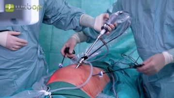
Trocar placement; Assessment of findings
![Trocar placement; Assessment of findings]()
Soundsettings Through the incision, the channel for the 10-mm trocar is first created with scissors and then inserted. Under diaphanoscopy and laparoscopic control with the camera, the two 5 mm working trocars are inserted in the right and left mid-abdomen respectively. The panoramic view shows clear medial hernia gaps in the lower abdomen on both sides.
Note 1: If no inguinal hernia is recognizable during inspection of the inguinal region, preparation should still be carried out, as the symptoms could be caused by the prolapse of a spermatic cord lipoma.
Note 2: After placing the trocars, the operating table is placed in the Trendelenburg position so that the intestines can be displaced to the upper abdomen, and tilted 20° toward the surgeon to enable better ergonomic working for him.
-
Opening of the Peritoneum on the Right; Dissection of the Preperitoneal Space
Soundsettings The incision of the peritoneum begins after external palpation in the area of the anterior superior iliac spine, runs in an arc 3-4 cm above the internal inguinal ring over the epigastric vessels and ends at the medial umbilical fold. The fold itself should not be severed.
Note: Asymptomatic adhesions in the lower abdomen do not need to be released, as the actual hernia repair is performed preperitoneally.
Medial to the epigastric vessels, preparation is then carried out between the bladder (caution: bladder injury) and the posterior wall of the rectus to the dorsal side of the symphysis and the Cooper's ligament is displayed.
Dissection of the right-sided hernia sac; Parietalization
Now, a larger medial hernia sac is dissected step by step (fascial defect M2 according to EHS (Euro
Now, a larger medial hernia sac is dissected step by step (fascial defect M2 according to EHS (Euro
Activate now and continue learning straight away.
Single Access
Activation of this course for 3 days.
US$9.10
inclusive VAT
Most popular offer
webop - Savings Flex
Combine our learning modules flexibly and save up to 50%.
from US$7.08 / module
US$85.05/ yearly payment
general and visceral surgery
Unlock all courses in this module.
US$14.17
/ month
US$170.10 / yearly payment
Webop is committed to education. That's why we offer all our content at a fair student rate.