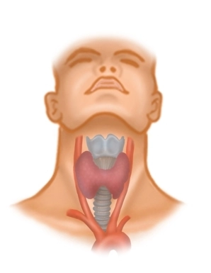
Situated between the anterior margin of the sternocleidomastoid muscle, the mandibula and the jugular fossa, the anterior cervical triangle in the vicinity of the hyoid bone comprises the suprahyoid and subhyoid muscles, vessels, nerves and the thyroid.
Fascial layers
The skin of the anterior triangle of the neck covers several fascial layers (all belonging to the cervical fascia) with distinctive features:
- The superficial lamina invests all structures of the neck, except for the platysma, and separately invests the sternocleidomastoid muscle as well as the posterior aspect of the trapezius muscle (accessory nerve XI).
- The medial pretracheal lamina invests the subhyoid muscles.
- The deep prevertebral lamina courses outside the surgical field between the esophagus and spine.
Just like the lateral vascular and nerve pedicle (carotid artery, internal jugular vein and vagus nerve), the trachea and thyroid / parathyroids also have their own organ fascias. With their three-dimensional configuration, the fascias invest compartments interspersed with spaces which extend into the mediastinum and thus represent potential routes of infection.