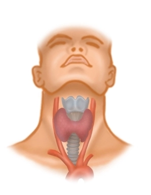
Located between the anterior border of the sternocleidomastoid muscle, the mandible, and the jugular fossa, the anterior triangle of the neck contains the supra- and infrahyoid muscles, vessels, nerves, and the thyroid gland in the vicinity of the hyoid bone.
Fascial Layers
In the anterior triangle of the neck, there are several fascial layers under the skin (all belonging to the cervical fascia), which exhibit certain peculiarities:
- The superficial lamina superficialis surrounds all neck structures except the platysma and separately encloses the sternocleidomastoid muscle and dorsally the trapezius muscle (accessory nerve XI).
- The middle lamina praetrachealis surrounds the infrahyoid muscles.
- The deep lamina praevertebralis runs between the esophagus and the spine outside the surgical area.
The trachea and the thyroid/parathyroid glands, as well as the lateral vascular-nerve bundle (carotid artery, internal jugular vein, and vagus nerve), also have their own organ fascias. The arrangement of the cervical fascias results in compartments enclosed by them and spaces (spatium) between them, which extend into the mediastinum and represent potential pathways for infection.