Start your free 3-day trial — no credit card required, full access included

Anatomy - Rectovaginal fistula: Anterior levatorplasty with simultaneous sphincter reconstruction

  1. Pelvic Floor

    Pelvic Floor

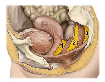
    (1) Rectogenital septum, (2) Paraproctium, (3) Mesorectal fascia

    The pelvic floor forms the muscular closure of the caudal abdominal cavity as a "functional unit" and prevents the prolapse of the intestines or pelvic organs. The pelvic floor structures support the intestinal and urogenital closure system, with the muscular components playing a crucial role in support and continence function. Muscular overstretching or denervation can lead to pelvic floor descent (descent) or to various forms of organ prolapse in the small pelvis. Women are much more frequently affected than men (gender ratio 9:1).
    The development of pelvic floor insufficiency is generally a multifactorial process. The most common causes of pelvic floor disorders include the number of vaginal deliveries, excessive straining during defecation, obesity, and previous surgical interventions in the small pelvis.

  2. Anal Canal (Canalis analis)

    Anal Canal (Canalis analis)

    (1) Longitudinal muscular layer, (2) Circular muscular layer, (3) Levator ani muscle, (4) Puborectalis muscle, (5) Deep external anal sphincter, (6) Anal columns, (7) Superficial external anal sphincter, (8) Subcutaneous external anal sphincter, (9) Kohlrausch's fold, (10) Internal sphincter, (11) Proctodeal gland, (12) Corrugator ani muscle

    Three muscles form the closure mechanism in the wall of the lower rectum:

    1. The internal anal sphincter is a thickening of the last circularly arranged fibers of the smooth colon musculature and is innervated by the sympathetic nervous system.
    2. The levator ani muscle, on the other hand, is voluntarily innervated (sacral plexus) and includes the puborectalis muscle attached to the pubic bone. It acts as a large sling around the anal canal, bending it functionally forward.
    3. The external anal sphincter is also striated and is suspended between the center of the perineal region (perineal body) and the coccyx. It is voluntarily innervated by the pudendal nerve. Its contraction closes the anal canal terminally. The different innervation of the three closing muscles provides additional protection against failures and resulting incontinence.

    In the mucosa of the anal canal, there are numerous longitudinal folds (anal columns) that have dense arterial plexuses with venous drainage. When the sphincter muscles contract, they quickly fill, and the mucosa swells, adheres together, and thus forms a gas-tight seal. Hemorrhoids and venous thromboses are known vascular complications in this region.
    Defecation occurs partly through the relaxation of the closure mechanisms (initiated by voluntary muscles, emptying of the erectile tissue) and partly through active abdominal pressure and intestinal peristalsis.

Rectum

Rectum (Rectum) Functionally, the rectum serves as a fecal reservoir, preventing continuous fecal d

Activate now and continue learning straight away.

Single Access

Activation of this course for 3 days.

US$9.20  inclusive VAT

Most popular offer

webop - Savings Flex

Combine our learning modules flexibly and save up to 50%.

from US$7.11 / module

US$85.43/ yearly payment

price overview

general and visceral surgery

Unlock all courses in this module.

US$14.23 / month

US$170.90 / yearly payment

to top