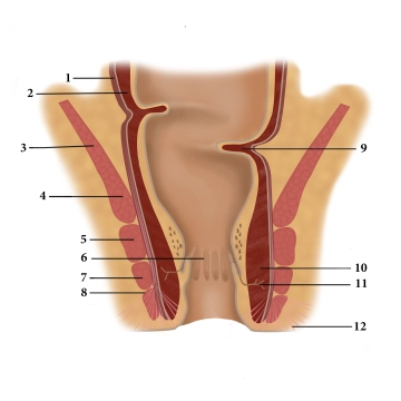
(1) Longitudinal muscular tunic, (2) Circular muscular tunic, (3) Levator ani muscle, (4) Puborectalis muscle, (5) Deep anal sphincter, (6) Anal columns, (7) Superficial anal sphincter, (8) Subcutaneous anal sphincter, (9) Kohlrausch fold, (10) Internal anal sphincter, (11) Proctodeal gland, (12) Corrugator ani muscle
Three muscles form the closure apparatus in the wall of the lower rectum:
- The internal anal sphincter is a thickening of the last circular fibers of the smooth colon muscle and is innervated by the sympathetic nervous system.
- The levator ani muscle, on the other hand, is voluntarily innervated (sacral plexus) and includes the puborectalis muscle attached to the pubic bone. It acts as a large sling around the anal canal, bending it functionally forward.
- The external anal sphincter is also striated and is suspended between the center of the perineal region (perineal body) and the coccyx. It is voluntarily innervated by the pudendal nerve. Its contraction closes the anal canal terminally.
The different innervation of the three closing muscles provides additional security against failures and resulting incontinence.
In the mucosa of the anal canal, there are numerous longitudinal folds (anal columns) that have dense arterial plexuses with venous drainage. Upon contraction of the sphincter muscles, they quickly fill and the mucosa swells, adheres together, and thus provides a gas-tight seal. Hemorrhoids and venous thromboses are known vascular complications of this region.
Defecation occurs partly through the relaxation of the closure mechanisms (initiated by voluntary muscles, emptying of the erectile tissue) and partly through active abdominal pressure and intestinal peristalsis.