Start your free 3-day trial β€” no credit card required, full access included

GIST - distal gastrectomy according to Roux-Y

Reading time readingtime 14:49 min.
  1. Preoperative Assessment

    Preoperative Assessment

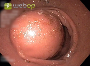
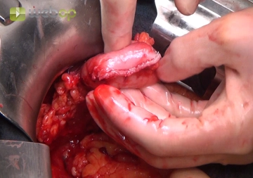
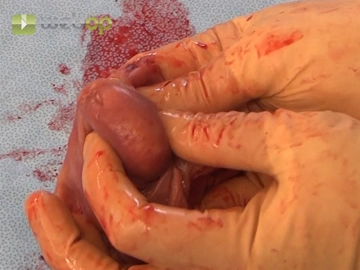
    The endoscopic examination reveals a submucosal tumor of the gastric wall obstructing the gastric outlet.

  2. Explorative Laparoscopy

    Video
    Explorative Laparoscopy
    Soundsettings

    The operation begins with a laparoscopic exploration of the abdomen to exclude peritoneal and liver metastases. In the course of this, an opportunistic cholecystectomy is performed independently of the tumor operation (not shown). After fenestration of the gastrocolic ligament with opening of the omental bursa, the posterior wall of the stomach becomes visible. The prepyloric tumor appears to be well resectable. Endosonographically, this tumor corresponds to an echo-heterogeneous 26 × 12 mm mass within the gastric wall.

  3. Conversion to Laparotomy

    Video
    Conversion to Laparotomy
    Soundsettings

    Due to the location of the tumor directly prepyloric, an atypical resection of the tumor is not possible, and there is an indication for distal gastrectomy. Therefore, conversion to an open procedure using a right-biased transverse upper abdominal laparotomy. After opening the abdominal cavity, the findings are inspected and palpated again.

  4. Mobilization of the Duodenum according to Kocher; Preparation of the Stomach

    Video
    Mobilization of the Duodenum according to Kocher; Preparation of the Stomach
    Soundsettings

    After mobilization of the descending part of the duodenum, the dissection of the proximal resection margin at the lesser and greater curvature is performed first. The preparation of the stomach can be done close to the stomach wall, as neither large safety margins nor lymph node dissection are necessary.

  5. Postpyloric Stomach Resection

    Video
    Postpyloric Stomach Resection
    Soundsettings

    Continue the dissection distally step by step at least 2 cm beyond the pylorus, severing the right gastric artery at the lesser curvature and the right gastroepiploic artery and vein near the gastric wall at the greater curvature. Once the duodenum is circumferentially mobilized postpylorically, it is transected with the stapling device.

  6. Oversewing of the Duodenal Stump

    Video
    Oversewing of the Duodenal Stump
    Soundsettings

    Oversewing the staple line of the duodenal stump with seromuscular interrupted sutures.

  7. Transection of the stomach proximally; Removal of the specimen

    Video
    Transection of the stomach proximally; Removal of the specimen
    Soundsettings

    The transection of the stomach is performed with a stapler, after which the specimen can be removed. Partial oversewing of the staple line with interrupted sutures, leaving a section approximately 5 cm long intended for the anastomosis.

  8. Formation of the Roux-Y Loop

    Video
    Formation of the Roux-Y Loop
    Soundsettings

    Approximately 30 cm aboral to the duodenojejunal flexure (Treitz's ligament), selection of a jejunal loop is performed under transillumination of the vascular arcades. Vascular-free portions of the mesentery are incised with the cautery, and the crossing vessels are divided between clamps. The intestine is transected with the stapler. The staple line of the efferent loop is oversewn in an inverting manner.

    The mesocolon is incised in a vascular-free region, and subsequently, the efferent jejunal loop is relocated retrocolically into the upper abdomen and approximated to the gastric stump. Care must be taken not to twist the mesentery.

  9. Gastrojejunostomy – Posterior Wall Suture

    Video
    Gastrojejunostomy – Posterior Wall Suture
    Soundsettings

    The anastomosis between the stomach and jejunum is performed end-to-side. The posterior wall suture is executed using a two-layer technique. After placing two lateral holding sutures, the anastomosis begins with a continuous seromuscular suture. Subsequently, the staple line in the area left free on the stomach is removed, and the opposite jejunum is opened to the same length. The second suture line is then completed as a continuous full-thickness suture with a monofilament thread.

  10. Gastrojejunostomy – Anterior Wall Suture

    Video
    Gastrojejunostomy – Anterior Wall Suture
    Soundsettings

    After stenting the anastomosis by advancing the gastric tube into the efferent jejunal limb, the anastomosis is completed with a single-layer suture using extramucosal interrupted sutures.

  11. Footpoint Anastomosis

    Video
    Footpoint Anastomosis
    Soundsettings

    The afferent jejunal loop is anastomosed end-to-side with the efferent jejunal loop 40 cm aboral to the gastrojejunostomy. Both loops are approximated with knotted corner sutures, followed by the resection of the staple line of the afferent loop and the antimesenteric opening of the afferent loop using an electrosurgical knife.
    The anastomosis technique corresponds to the technique shown in steps 9 and 10 for the gastrojejunostomy. It is performed in two layers in the area of the posterior wall and as a single-layer extramucosal interrupted suture in the area of the anterior wall.

  12. Abdominal Wall Closure

    Video
    Soundsettings

    After placing a drain at the duodenal stump, a two-layer fascial closure is performed, along with a subcutaneous suture. The skin suture is performed as an intradermal suture with absorbable thread.

Adverts

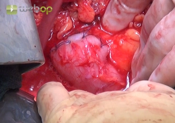
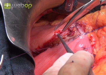
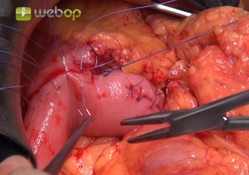
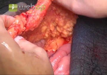
Active local hemostasis and sealing

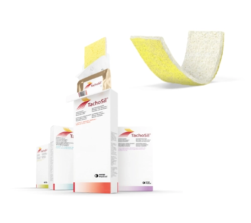
TachoSil® Versiegelungsmatrix

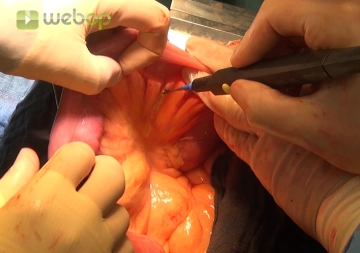
TachoSil® is used in adults and children from 1 month of age as supportive treatment in surgery for improving hemostasis, for supporting tissue sealing, and for suture support in vascular surgery when standard techniques are insufficient. TachoSil® is used in adults for supportive sealing of the dura mater to prevent postoperative cerebrospinal fluid leakage after neurosurgical procedures.

Produktwebsite TachoSil®
TachoSil® Prescribing Information 05-2025 (354.1 kB)

to top