Anatomy - pelvic lymphadenectomy on both sides, laparoscopic, robot-assisted laparoscopy (DaVinci)

  1. The bony pelvis

    The bony pelvis

    The human pelvis is a complex ring structure that fulfills two main functions. On one hand, it forms the lower closure of the trunk and thus carries the weight of the upper body. On the other hand, it transfers this weight to the lower limbs, which is crucial for the upright posture and locomotion of humans.

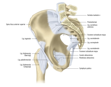
    The pelvis is composed of several bony elements. The foundation is formed by the two hip bones, also known as the pelvic girdle or cingulum membri inferioris. Between them is the sacrum. The os coxae consists of three main components:

    1. The ilium (Os ilium), consisting of the corpus ossis ilii and the ala ossis ilii.
    2. The ischium (Os ischii), consisting of the corpus ossis ischii and the ramus ossis ischii.
    3. The pubis (Os pubis), consisting of the corpus ossis pubis, the ramus superior, and the ramus inferior ossis pubis.

    These bones and the sacrum are connected by various types of connections, including ligamentous connections, cartilaginous connections, and joints:

    1. Ligamentous connections include the membrana obturatoria, the ligamenta sacroiliaca, sacrotuberale, and sacrospinale.
    2. Fibrocartilaginous connections are established by the symphysis pubica.
    3. Joint connections include the articulation sacroiliaca and the articulatio sacrococcygea.

    There are various ligaments involved in the joints:

    • The ligg. sacroiliaca anteriora and posteriora are ligaments that connect the upper and lower ends of the ilium to the sacrum.
    • The ligg. sacroiliaca interossea extend between the sacrum and the tuberositas ossis sacri and tuberositas iliaca.
    • The lig. iliolumbale runs from the 4th and 5th lumbar vertebrae to the crista iliaca and the tuberositas iliaca.
    • The ligg. sacrotuberale et sacrospinale are additional ligaments that act on the articulatio sacroiliaca.
    • The lig. sacrococcygeum anterius, the lig. sacrococcygeum posterius profundum, and the lig. sacrococcygeum posterius superficiale are ligaments that connect the sacrum to the coccyx.

    The pelvis is divided into two main parts:

    • the greater pelvis (pelvis major) and
    • the lesser pelvis (pelvis minor).

    The linea terminalis is the dividing line between them. The greater pelvis, above the linea terminalis, contains the space between the two iliac wings, while the lesser pelvis includes the pelvic inlet, the pelvic outlet, and the pelvic walls.

    The pelvic walls are covered by connective tissue, known as fasciae. These fasciae are important for the stability and delineation of the pelvic organs. Furthermore, there are openings in the pelvic walls that allow the passage of blood vessels, nerves, and muscles that run between the pelvis and the buttocks. The pelvic region is rich in neurovascular structures that play an important role in supplying the pelvis and lower extremities.
  2. Pelvic diaphragm

    Pelvic diaphragm 1
    Pelvic diaphragm 2

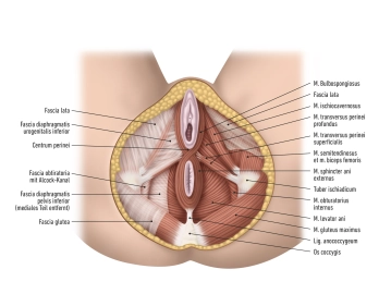
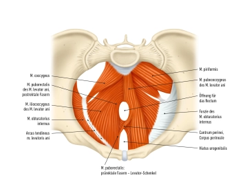
    The pelvic diaphragm is a term that describes the muscular structure forming the pelvic floor. It consists of three main muscles:

    M. levator ani: The muscle forms a funnel-like structure with an open tip downward. On its front side, the urogenital hiatus opens, a gap extending from the posterior side of the pubic bone to the perineal center. The M. levator ani frames this hiatus with its two limbs, which unite in front of the rectum at the perineal center. This center forms the muscular and connective tissue foundation of the perineum. The urogenital hiatus itself is covered and closed by the urogenital diaphragm. The levator ani is composed of the following muscles:

    • M. puborectalis,
    • M. pubococcygeus
    • M. iliococcygeus.

    M. coccygeus: This muscle extends from the ischial spine to the coccyx and runs like the M. levator ani in the pelvic floor.

    M. sphincter ani externus: This is the external anal sphincter and consists of striated muscle tissue. It has three parts: Pars subcutanea, Pars superficialis, and Pars profunda. The Pars subcutanea consists of superficial fibers running in front of and behind the anus. The Pars superficialis forms muscle plates on the sides of the rectum and acts like a clamp. The Pars profunda consists of circularly arranged muscle fibers surrounding the rectum, enabling voluntary closure of the bowel.

    The pelvic floor also contains the urogenital diaphragm, which closes the urogenital hiatus. It consists of various muscles located in the superficial perineal space (superficial pelvic floor space) and the deep perineal space (deep pelvic floor space):

    M. transversus perinei superficialis: a flat, transverse muscle of the pelvic floor. It belongs to the superficial perineal muscles and extends from the pubic bone (Ramus inferior ossis pubis) to the center of the perineum, where it merges with the muscle of the opposite side or connects with the fibers of the external anal sphincter.

    M. transversus perinei profundus: It lies below the M. transversus perinei superficialis and is stronger and more deeply embedded in the structure of the pelvic floor compared to it. The muscle also runs transversely from the inner edge of the ischium (Ramus inferior ossis ischii) to the midline of the body, where it meets the muscle of the opposite side or connects with the fibers of the external sphincter of the urethra and partially the M. sphincter ani externus.

    M. sphincter urethrae externus: This is the external sphincter of the urethra and is formed by the M. transversus perinei profundus.

    Additionally, there are two muscles adjacent to the internal genital organs:

    M. ischiocavernosus: This muscle attaches to the ischial rami and covers the clitoral body. It is involved in erection.

    M. bulbospongiosus (M. bulbocavernosus): This muscle runs from the perineal center and covers the vestibular gland. In women, it can narrow the vestibule of the vagina.

    The pelvic fascia, also known as the pelvic fascia, plays a crucial role in the structure and function of the pelvis. It encompasses a series of connective tissue layers that support the pelvic organs, envelop the musculature, and contribute to the overall stability of the pelvic floor. This fascial structure is divided into two main parts: the parietal pelvic fascia and the visceral pelvic fascia.

    1. Parietal pelvic fascia (Fascia endopelvina) This fascial layer lines the inner walls of the pelvis. It covers the muscles forming the pelvis, including the M. piriformis and the M. obturator internus. The parietal pelvic fascia contributes to the demarcation of pelvic spaces and forms important anatomical structures such as the retropubic space and the ischioanal space. It also provides an attachment surface for other fasciae and ligaments that hold the pelvic organs in position.
    2. Visceral pelvic fascia (Fascia propria organi): The visceral pelvic fascia encloses the pelvic organs and adapts to their shape changes. Each organ is surrounded by a specific partial fascia, giving it a unique name. Additionally, this fascia forms various septa that separate the organs, including the vesicovaginal septum between the bladder and vagina and the rectovaginal septum between the rectum and vagina. The accompanying connective tissue, which carries blood vessels and nerves to the respective organs, is specifically named: as Paraproctium, supporting the rectum, as Paracystium, supporting the bladder, as Parametrium, surrounding the uterus, and as Paracolpium, structuring the vagina.

    There are also openings in the wall of the lesser pelvis that allow the passage of nerves and blood vessels:

    Obturator canal: A canal for the obturator nerve and vessels, running from the lesser pelvis to the inner thigh.

    Ischiadic foramina: These allow the passage of vessels, nerves, and muscles from the lesser pelvis into the gluteal region. There is the greater sciatic foramen and the lesser sciatic foramen.

    Finally, the lesser pelvis houses internal genital organs, the bladder, part of the ureter, and the rectum. These organs are surrounded by fat and connective tissue, and vessels and nerves run in the pelvic wall and connective tissue to supply and innervate them.

  3. Rectum

    • Origin of the Rectum: Extension of the sigmoid colon, beginning at the upper edge of the third sacral vertebra.
    • Shape and Curvatures:
      • Sacral flexure: Follows the contour of the sacrum downward.
      • Anorectal (perineal) flexure: Bends backward in the perineal region, bulging forward.
      • Lateral flexures: Occasionally variable curvatures in the frontal plane.
    • Structural Features:
      • Length of approximately 15 cm.
      • Three semilunar transverse folds; the most prominent is the Kohlrausch fold about 6 cm above the anus on the right side.
    • Continuation into the Anal Canal: Ampulla recti above the anal canal, which expands when filled.
    • Boundaries and Position:
      • Upper section: Covered by peritoneum in front and laterally, retroperitoneal (Rectum fixum).
      • Peritoneal contact: In women, the peritoneum meets the posterior vaginal fornix and the back of the uterus (rectouterine excavation) at the middle transverse fold.
      • Below the Kohlrausch fold: Completely extraperitoneal.
    • Anatomical Neighborhood:
      • Adjacent to the sacrum, coccyx, and the posterior parts of the levator ani muscle.
      • Bordering the vagina in front.
    • Vasculature and Innervation:
      • Median sacral artery, superior rectal artery.
      • Sympathetic trunks, lateral sacral arteries, sacral plexus in retrorectal connective tissue.
  4. Ureter

    Pelvic and intramural part of the ureter: Section extending from the crossing of the linea terminalis with the sacroiliac joint to the entrance into the bladder wall.

    • Course of the ureter: On the right, the ureter runs above the initial segment of the external iliac artery and on the left over the bifurcation of the common iliac artery.
    • Peritoneal coverage: Initially covered by peritoneum, the ureter follows the lateral wall of the small pelvis, positioning itself ventral to the internal iliac artery.
    • Change in direction of the ureter: Near the ischial spine, the ureter turns frontal and medial towards the bladder, separating from the peritoneum.
    • Crossing with the broad ligament of the uterus: The ureter crosses this ligament, running below the uterine artery and near the lateral vaginal fornix.
    • Intramural part: This part of the ureter obliquely penetrates the bladder wall and opens into the bladder at the ureteral orifice.
  5. Urinary bladder

    The urinary bladder is a muscular hollow organ that serves to store urine and later expel it through the urethra. The bladder's capacity is normally 300-500 ml. The bladder can be divided into different parts,

    • the bladder apex (Apex vesicae), 
    • the bladder body (Corpus vesicae), 
    • bladder base (Fundus vesicae), 
    • bladder neck (Collum or Cervix vesicae), transition to the urethra 

    The apex is connected to the median umbilical ligament (Chorda urachi), the obliterated allantoic duct leading to the navel. 

    The peritoneum extends from the bladder to the uterus (Excavatio vesicouterina).

    The trigone of the bladder is a roughly triangular, smooth mucosal area located at the base of the urinary bladder. This triangle is formed by the two openings of the ureters (Ostia ureterum) and the internal opening of the urethra (Ostium urethrae internum). The interureteric fold bounds the trigone posteriorly, and anteriorly it forms an angle that extends into the urethra. In this area, the tunica mucosa is closely fused with the tunica muscularis. In other areas of the bladder, there exists a tunica submucosa, which forms a slightly movable buffer zone between the mucosa and muscle layer, allowing the bladder to fill and empty without difficulty.  

Urethra

The female urethra is approximately 3-5 centimeters long. It is anteriorly connected to the connect

Activate now and continue learning straight away.

Single Access

Activation of this course for 3 days.

US$9.10  inclusive VAT

Most popular offer

webop - Savings Flex

Combine our learning modules flexibly and save up to 50%.

from US$4.20 / module

US$50.48/ yearly payment

price overview

gynecology

Unlock all courses in this module.

US$8.41 / month

US$101.00 / yearly payment